DSS: Redefining Biotechnology & Life Science in India


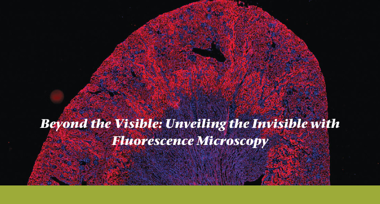
Beyond the Visible: Unveiling the Invisible with Fluorescence Microscopy
Beyond the Visible: Unveiling the Invisible with Fluorescence Microscopy
The phenomenon of fluorescence, with its mesmerizing dance between absorption and emission, allows us to see beyond the limits of natural vision, harnessing the power of illumination to transform scientific exploration. Behind the lens of a fluorescence microscope, scientists become storytellers, capturing these luminescent tales through careful observation and precise instrumentation. Every detail is meticulously documented, each image an iridescent masterpiece that reveals the intricate choreography of cellular life.
Through this blog, we invite you to embark on a thrilling expedition into the captivating world of fluorescence microscopy.
History of Fluorescence Microscopy
When researchers realized that some compounds, including quinine, fluoresced when exposed to ultraviolet (UV) light in the early 20th century, fluorescence microscopy was born. In the 1920s, German researcher August Köhler used fluorescence for the first time to examine biological material.
The field of fluorescence microscopy revolutionized in the 1950s when fluorescent dyes were used to designate biological material. George Wald, an American physicist, won the 1967 Nobel Prize in Physiology or Medicine for his groundbreaking work using fluorescence microscopy to study the visual pigments in the eye.
The resolution and sensitivity of fluorescence microscopy were significantly enhanced in the 1970s by creating fresh fluorophores and cutting-edge imaging methods like confocal and two-photon microscopy. This gave birth to modern-day fluorescence microscopy, making it an essential tool in biological research.
Working of Fluorescence Microscopy
A material emits light of a longer wavelength (lower energy) after absorbing light of a shorter wavelength (higher energy), and this is the basis for fluorescence, which is the concept upon which fluorescence microscopy is based.
Below is a detailed description of how fluorescence microscopy operates:
- Fluorophore labeling: Fluorophores are specialized fluorescent compounds that are often used to mark the sample of interest, such as cells or tissues. These fluorophores can bind to specific molecules in the sample or be directly linked to the target molecules of interest, such as proteins or DNA.
- Filter system: The excitation light travels through a number of filters that only allow the desired wavelengths of light to pass through. Only the excitation wavelength may reach the sample due to a filter in the excitation light route, which blocks all other wavelengths. By doing this, background noise is reduced and fluorophores are excited to their greatest potential.
- Excitation: A light source, usually a high-intensity lamp or a laser, is used to illuminate the sample in order to excite the fluorophores. The excitation light is chosen such that its wavelength is shorter than the fluorescence light that is produced. For instance, the fluorophore may be stimulated by blue light if it emits green light.
- Emission: Fluorescence emission occurs when excited electrons within fluorophores release extra energy after absorbing it. This allows the excited electrons to transition back to their ground state. This light is then released, and it may be of a different color and have a longer wavelength than the excitation light.
- Second filters: Another set of filters is employed to specifically identify the fluorescence light that is emitting while blocking the excitation light. Just the fluorescence emission wavelength can pass through a barrier filter in the emission light channel; excitation light and other undesired wavelengths are blocked.
- Formation of image: The objective lens of the microscope receives the emitted fluorescence light after it has passed through the filter system. The sample is illuminated by the objective lens, which also gathers the fluorescence signals that are released. Then, after being enlarged, these signals are directed onto a detector, such as a camera or a photomultiplier tube (PMT).
Fluorescence microscopy applications in biological research
Molecular tracking and visualization within living cells and tissues are now possible because of fluorescence microscopy, which has completely changed biological research. Fluorescence microscopy is widely used in the analysis of cellular organization and structure. Researchers can see the spatial distribution and dynamics of particular organelles or cellular components within the cell by labeling them using fluorescent probes. This makes it possible to study the connections between organelles, intracellular trafficking, and the development of intricate cellular structures.
Fluorescence microscopy also plays a crucial role in elucidating protein function and dynamics. Through the use of fluorescent protein tags or antibody labeling, researchers can track the localization, movement, and behavior of individual proteins in real time. This enables the study of protein-protein interactions, post-translational modifications, and protein turnover within living cells. Additionally, fluorescence resonance energy transfer (FRET) techniques can be employed to investigate protein conformational changes and signaling events, providing a deeper understanding of protein function and regulation.
Additionally, fluorescence microscopy is becoming a crucial technique in the study of immunity. Researchers may see and analyze immune responses, recognize certain immune cell subsets, and investigate immune cell interactions inside tissues by fluorescently labeling antibodies or immune cells. This makes it easier to comprehend how the immune system affects inflammation, autoimmune illnesses, and infections, which will help novel treatment approaches be created.
In biological studies, fluorescence microscopy Accessories has become a crucial technique. It has opened up new perspectives for comprehending the underlying biological processes because of its capacity to detect and monitor molecules within living cells and tissues.
The benefits and drawbacks of fluorescent microscopy
Here are some advantages and disadvantages of fluorescence microscopy to consider:
Advantages:
Specificity: The great specificity of fluorescence microscopy is one of its key benefits. Researchers can selectively view and analyze particular components inside a sample by utilizing fluorescent probes or antibodies that bind to target molecules individually. This makes it possible to precisely identify and analyze relevant chemicals.
Sensitivity: Fluorescence microscopy has a high sensitivity that enables the identification of fluorescently labeled molecules at low concentrations. This is very useful when researching uncommon or sparse targets in intricate biological samples.
Non-invasive and Live-cell Imaging: Live cells may be imaged using fluorescence microscopy without compromising their viability or functioning. This non-invasive nature enables the study of dynamic phenomena in their natural environments by allowing researchers to observe and track cellular activities over long periods of time.
Disadvantages:
Photobleaching is the irreversible reduction of fluorescence intensity that occurs over time as a result of exposure to the excitation light, and it can affect fluorophores used in fluorescence microscopy. Due to this, imaging tests may only last so long, and the imaging settings must be carefully optimized to reduce photobleaching.
Phototoxicity: Constant exposure to excitation light may result in cellular damage, which may alter cell viability and behavior. To reduce phototoxic effects, researchers must improve imaging setups and reduce light intensity and exposure time.
Background Noise: Autofluorescence from the sample or nonspecific fluorophore binding are examples of background noise that may be encountered during fluorescence microscopy. The accuracy of the results may be impacted and the signal-to-noise ratio may decrease due to certain sources of noise.
Upcoming Fluorescence Microscopy Developments
Fluorescence microscopy continues to evolve and advance, driven by ongoing research and technological innovations. Some upcoming exciting developments in this field are:
Multimodal Imaging: Fluorescence microscopy may be combined with other imaging techniques like electron microscopy (EM) or light-sheet microscopy to provide a more complete picture of biological processes and structures.
Single-Molecule Imaging: Using single-molecule imaging methods, it is possible to identify and follow specific fluorescently marked molecules inside live cells.
In vivo and Deep Tissue Imaging: Research is ongoing to find ways to image thick tissues or whole organisms without being constrained by fluorescence microscopy. Deeper tissue penetration is made possible by methods like light-sheet microscopy and multiphoton microscopy, which also reduce scattering and increase imaging depth.
Fluorescence microscopy will likely be crucial in pushing the limits of scientific investigation at the microscopic level as technology develops further.
Latest Articles

The Evolution of Customer Support in Clinical Diagnostics
BY Surender Kumar | General Manager – Service, Clinical Diagnostics Division 23rd July 2026
How Customer Expectations Have Changed Over the Last 20 Years “Customer Support is no longer a cost center—it is a competitive advantage.” Twenty years ago, customer support was primarily viewed...
Read More
The ROI of Lab Automation: How Automated Karyotyping Reduces Costs...
BY DSS Imagetech 13th July 2026
If you walk into almost any cytogenetics lab across India right now, you’ll probably hear the exact same complaint. Lab directors are staring at their monthly numbers, calculating just how...
Read More
Journey from Manual Testing to Automation powered by AI &...
BY Mr. Mahesh Semwal, Deputy General Manager (DGM) 29th June 2026
The diagnostics industry has witnessed a tremendous transformation over the last 25–30 years. Those who have been part of this journey have seen how diagnostics laboratories have moved from intensive...
Read More