DSS: Redefining Biotechnology & Life Science in India


Home > Products & Services > Instruments > H&E
H&E
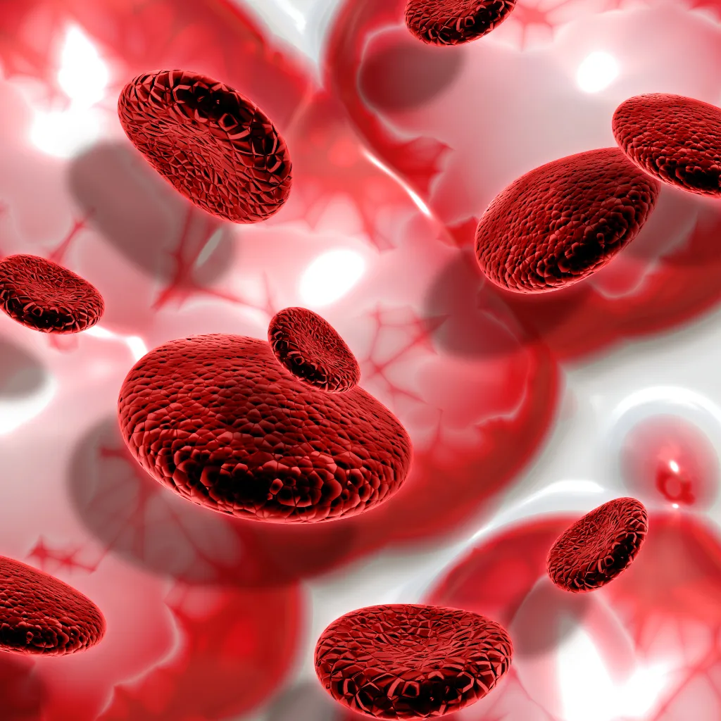
A combination of two hematoxylin stains- hematoxylin and eosin make H&E. The hematoxylin stains cell nuclei blue, and eosin stains the extracellular matrix and cytoplasm pink, with different designs taking on various shades, hues, and mixes of these colours. For regular diagnosis, the method of Hematoxylin and Eosin (H&E) is by far favoured for observing cellular and tissue structure detail by pathologists. The variety of stain energy is regularly determined by the pathologist’s learning experience and personal preference. Since this stain shows a particularly wide scope of cytoplasmic, nuclear, and extracellular matrix features, virtually all teaching texts use H&E pictures. This simple and essential stain is used even today, the process being unchanged for over a century.
Biopsy specimens are grossed and proccesed to produce Tissue sections which are Universally stained with Hematoxylin & Eosin to make them visible by Microscope. Automated H&E stainer makes this process consistent and faster, with optimised staining Protocol.
DSS has been representing the H&E solution combines the advanced Dako CoverStainer with ready-to-use reagents, a validated, optimized protocol, and features – an intelligent, automated reagent handling system.
What are Hematoxylin and Eosin staining used for in laboratory settings?
Hematoxylin and Eosin (H&E) staining is widely used in laboratory settings, particularly in histology, pathology, and biology. It is a staining method commonly used to prepare and examine tissue samples under a microscope.
The H&E staining process involves a sequence of steps, including tissue fixation, dehydration, embedding in paraffin, sectioning into thin slices, staining with hematoxylin and eosin dyes, and mounting on slides. After staining, the tissue sections can be examined under a light microscope to gain insights into the tissue’s cellular and structural composition. It is a cornerstone of histopathology and is essential for diagnosing diseases, understanding tissue biology, and advancing medical research.
DSS Imagetech has been serving as the representative for the H&E solution, a comprehensive offering that integrates the cutting-edge Dako CoverStainer with pre-prepared reagents, a rigorously validated and optimized protocol, and an array of intelligent, automated reagent handling features.
How does H&E staining help in histopathology?
DSS Imagetech represents Dako H&E staining methods commonly used in histopathology and plays a crucial role in the field of diagnostic medicine and biomedical research.
H&E staining is an essential technique in histopathology that enables the examination, diagnosis, and research of tissue samples. It enhances the visibility of cellular and structural details, aids in disease identification, and supports both clinical and research endeavours in the field of medicine and biology.
Are there different staining protocols for Hematoxylin and Eosin staining?
DSS Imagetech H&E staining remains the same, there can be variations in staining protocols and techniques, which may be influenced by factors such as the type of tissue, the specific laboratory’s standard operating procedures, and the desired staining intensity.
the basic H&E staining steps are consistent, variations may exist in the specific formulations of hematoxylin and eosin solutions, staining times, and the use of counterstains.
It’s essential to follow established protocols and adapt them as needed to achieve the desired staining results for different types of tissues and research objectives.
What are the key steps in the H&E staining process?
Hematoxylin and Eosin (H&E) staining is a widely used technique in histology and pathology to visualize the microscopic structure of tissue samples. It helps distinguish various cell types and tissue components by staining cell nuclei (with hematoxylin) and cytoplasm and other structures (with eosin).
The key steps in the H&E staining process are Tissue preparation, Embedding, Sectioning, Deparaffinization, Rehydration, Staining, Dehydration and Clearing, Mounting, Drying and Microscopic examination.
DSS Imagetech H&E staining is a technique in histopathology and provides valuable information for diagnosing diseases and understanding tissue morphology.
Can H&E staining be used on various tissue types?
Yes, Hematoxylin and Eosin (H&E) staining is a widely used histological staining technique that can be applied to a variety of tissue types. H&E staining is commonly used in the field of histology to visualize the microscopic structures and cellular details of tissues. It provides good contrast and allows for the differentiation of different tissue components.
What are the typical challenges associated with H&E staining?
Hematoxylin and eosin (H&E) staining is a widely used histological technique in pathology and research to visualize the cellular and tissue structures of biological specimens. While it is a valuable tool, there are several challenges associated with H&E staining: Tissue Processing Variability, Artifacts, Staining Variability, Tissue Over- or Under-Staining, Nuclear Detail, Counterstaining, Tissue Specificity, Microscope Variation etc.
Despite these challenges, DSS Imagetech H&E staining remains a valuable tool for histological analysis, and with proper technique and attention to detail, many of these issues can be minimized or overcome. Researchers and pathologists often employ various quality control measures to ensure reliable staining results and accurate interpretation of tissue specimens.
How long does a Hematoxylin and Eosin staining procedure usually take?
The duration of a Hematoxylin and Eosin (H&E) staining procedure can vary depending on several factors, including the specific protocol or laboratory practices, the type of tissue or specimen being stained, and the equipment used. However, a typical H&E staining procedure can take anywhere from 1 to 2 hours to complete.
Is there any special equipment required for H&E staining?
Hematoxylin and Eosin (H&E) staining is a widely used technique in histology and pathology to visualize tissue samples under a microscope. While it doesn’t require highly specialized equipment, it does involve several key pieces of equipment and chemicals
What are the differences between Hematoxylin and Eosin staining and other histological stains?
Hematoxylin and Eosin (H&E) staining, a widely utilized histological procedure, imparts distinct colors to cellular components. Hematoxylin stains nuclei and nuclear components with a spectrum of blue-purple shades, while Eosin stains varied pink shades to extracellular components and cytoplasm. H&E stains are prevalent in routine histopathology due to their rapid and cost-effective nature, distinguishing them from other stains like Periodic acid-Schiff (PAS), Masson’s trichrome, Orcein, Reticulin, and Gomori’s Methenamine-Silver (GMS), each tailored for specific purposes, tissue types, cellular structures etc.
Biopsy specimens undergo grossing to produce universally stained tissue sections with Hematoxylin & Eosin, rendering them visible under a microscope. The automation of this process, facilitated by the Dako Coverstainer developed by Dako and distributed by DSS in India, enhances consistency and expedites the staining protocol. This system, characterized by advanced technology, alleviates the laborious aspects of the procedure for pathologists, seamlessly integrating automation from the initial stages of baking to the final drying phase.
Can H&E staining be automated for high-throughput applications?
Yes, Dako H&E (Hematoxylin and Eosin) staining represented by DSS Imagetech can be automated for high-throughput applications. Automation of H&E staining processes has become increasingly common in research, clinical, and diagnostic laboratories to improve efficiency, consistency, and throughput. Here are some of the ways H&E staining can be automated:
Automated Stainers: Specialized automated staining machines are available that can perform the entire H&E staining process.
Slide Loaders: Automated slide loaders can load multiple slides onto staining racks, streamlining the sample handling process.
Automated Cover Slips: After staining, an automated cover slipping system can be used to apply a coverslip to the slides, reducing the need for manual intervention.
Digital Pathology: Digital pathology systems can capture high-resolution images of stained slides and automate the process of digitizing and analyzing tissue samples.
It’s important to note that the specific automation methods and equipment used can vary depending on the laboratory’s needs and resources. The choice of automation system should be based on factors like sample volume, budget, and the level of automation required.
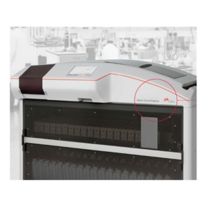
Dako CoverStainer
Dako CoverStainer automates every step of the primary staining process from baking to drying, streamlining workflow and providing consistent quality slide after slide.
Testimonials & Reviews
Dr. (Prof.), Nitesh Mohan
Professor & Head, Department of Pathology, RMCH Bareilly
DSS's expertise, dedication, and professionalism were outstanding in making the Karyotyping & FISH workshop a great success. Their knowledge and valuable insights empowered all the participants with practical skills, receiving highly positive feedback from both students as well as faculty members.
Dr. Chhaya Chande, Professor & HOD, Microbiology
GGMCJJ Hospitals, Mumbai
“Ms. Megha Dhumal (Assistant Manager- Application) has done a satisfactory demonstration of the running of the Abbott Sample preparation machine model m2000sp and the Abbott RT-PCR machine model m2000rt. We appreciate the effort made by the DSS team under these difficult conditions to help our lab to carry out the imperative Covid-19 tests.”
Dr Sunil K Arora, Professor, Deptt of Immunopathology
PGIMER, Chandigarh
“We are using Confocal Microscope and one Fluorescence Microscope. Both are working fine. The after sales services by DSS have been excellent for functioning & upkeep of the microscopes. The applications support by experts from DSS is very useful. Keep it up!”
Dr Pramod Kumar Bajaj
MD, Spermprocessor Pvt Ltd
“Really excited to see the DSS Pathology solutions exhibition booth at APCON 2019 along with Magnus. We think all the upcoming technology had been displayed along with your efforts to make it Indigenous (Made in India) is highly appreciated. Wish you all the best. Keep it up!”
Dr. Sreejesh S, Associate Professor, Dept of Hematology
PGIMER, Chandigarh
“My experience with DSS so far has been very good till now. We are getting good support in both purchase as well as in troubleshooting. Very good experience with Mr Arun, Mr Manoj, Mr Mahesh and all others from the DSS team.”
Dr Sudha S Murthy, Department of Pathology and Laboratory Medicine
BIACH & RI, Hyderabad
“I am happy with DSS and associated with 19 years and use Dako antibody. Happy with Supply but need improvement.”
Dr S Radhika MD, PhD
Professor, Deptt. Of Cytology & Gynaec Pathology, PGIMER, Chandigarh
“PGI Cytology Dept. has had a long association with DSS- Olympus Microscopy Division. They have provided excellent services- after sales service. The product is also of very good quality. We have had no problems with their products and services are of very good quality.”
Dr Nuzhat Husain
RMLIMS, Lucknow
“Have been using Dako Reagents and Dako antibodies for a while. Services and products have been good and timely.”
Dr Minu Singh
Assistant Professor, PGIMER, Chandigarh
“MRC Holland MLPA products provided by DSS are of good quality, have never faced any quality issues with their product or shipping condition. They provide prompt response upon any query.”
Mr. Krishnani Professor, SGPGI, Lucknow
“My experience with DSS so far has been excellent for the last 30 years- sales and service experience. Microscope products are very useful and sturdy with high precision.”