DSS: Redefining Biotechnology & Life Science in India



























Home > Applications & Specialities > Microscopy
Microscopy
A microscope is an instrument that magnifies objects, too small to be seen with an unaided eye, producing larger images using a combination of lenses. Student lab microscopes tend to be brightfield microscopes, meaning that visible light is passed through the sample and used to form an image directly, without any modifications. Another type of light microscopy is fluorescence microscopy, which is used to image samples that fluoresce (absorb one wavelength of light and emit another). For further improved resolution, Confocal Microscopy is used. A confocal microscope uses a laser to excite a thin layer of the sample and collects only the emitted light coming from the target layer, producing a sharp image without interference from fluorescent molecules in the surrounding layer. Selection of the type microscopes is based on the type of sample (size, thickness, dynamics, physiology etc.) need to be imaged.


Confocal
Seeing is believing, a well-known phrase holds excellent significance in science and in this context, optical microscopy is an important,…
Light Sheet
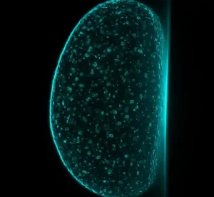
At DSS Imagetech, we bring cutting-edge imaging solutions to the forefront of scientific research. Light Sheet Microscopy (LSM), also known…

Scientific Imaging
Scientific imaging refers to the acquisition, processing, and analysis of images in a scientific context. In the field of microscopy,…
Super Resolution
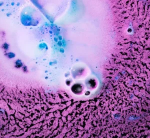
Fluorescence microscopy, with its major impact on biomedical research over past several decades, the resolution barrier (Diffraction limit) inherent in…

Widefield
At DSS Imagetech, we are committed to delivering precision and innovation in microscopy solutions. Our Wide Field Microscopy systems are…
Microscopy Applications for Life Science Research
Every microscopy project starts with a question, not a technique: what do you need to see, and what will happen to the sample while you look? That question is what separates the microscopy applications on this page.
A thin fixed slide, a living cell over several hours, or a thick three-dimensional tissue sample each call for a different approach to image acquisition. Scientific imaging as a whole depends on matching the technique to the specimen, rather than the other way round.
Understanding Microscopy in Life Sciences
Microscopy lets researchers observe and analyse structures and processes that are invisible to the naked eye, from whole cells down to sub-cellular detail. What a researcher can actually extract from that observation depends on four things working together: how the sample is prepared, how contrast is generated, how the image is acquired, and how it's subsequently analysed.
Get any one of these wrong and the imaging technique itself won't compensate.
Selecting an imaging approach should start with the biological question, not the instrument. A question about protein localisation within a thin cell layer points toward a different technique than a question about how a structure changes shape over hours inside living tissue.
Four properties tend to shape that decision most directly: spatial resolution (how much fine detail the image can resolve), imaging depth (how far into a thick or three-dimensional sample the technique can image clearly), field of view (how much of the sample is captured at once), and acquisition speed (how quickly images can be captured, which matters when a process is moving or changing).
Research microscopy usually involves trading off between these properties rather than maximising all four at once.
Explore Advanced Microscopy Techniques
DSS Imagetech's microscopy applications hub covers five advanced microscopy techniques currently represented in the portfolio: widefield, confocal, light-sheet, super-resolution and scientific imaging.
Each technique supports different combinations of sample type, research question and imaging workflow, so selection depends on the specific experimental requirement.
Widefield Microscopy
Widefield microscopy illuminates and captures the entire field of view simultaneously, rather than scanning point by point. It's commonly used for routine fluorescence and transmitted-light imaging, where a full picture of the sample is needed quickly.
Widefield approaches suit thinner samples and applications such as general cell observation, where speed and simplicity matter more than optical sectioning.
The main limitation is that out-of-focus light from other planes in a thicker sample can blur the in-focus image, which is one reason other techniques exist for thicker specimens. Explore the DSS Imagetech widefield microscopy category for more detail.
Confocal Microscopy
A pinhole is doing the real work here: it strips out light from every plane except the one in focus, so confocal microscopy only collects signals from a single thin optical slice. Image enough of those slices and you can reconstruct a specimen that would otherwise be too thick to image clearly in one pass — that's optical sectioning.
It matters most on fluorescently labelled samples and general cellular imaging, anywhere a widefield shot would just come back with the in-focus structure buried under blur from everything around it.
The consideration to weigh is acquisition speed and, in some configurations, phototoxicity from the laser illumination used to generate the optical section — factors worth checking against your specific sample and experiment duration. See DSS Imagetech's confocal microscopy category for further reading.
Light-Sheet Microscopy
Instead of flooding the whole specimen with light, light-sheet microscopy illuminates one thin plane at a time — which is how it builds a full volumetric image without exposing everything else in the sample to unnecessary light.
That's the whole reason it shows up so often in developmental biology and extended live-sample imaging: phototoxicity and photobleaching stop being a problem when only a sliver of the specimen is lit at any given moment.
Sample mounting and optical geometry are fairly specific for these systems, though, so it's worth confirming compatibility with your setup before committing to it. Explore the DSS Imagetech light-sheet microscopy category to learn more.
Super-Resolution Microscopy
Super-resolution microscopy extends imaging capability beyond the conventional diffraction-limited resolution of light microscopy, allowing researchers to resolve structural detail that would otherwise blur together under standard optical microscopy.
It's generally considered for subcellular and molecular-scale structural questions where standard resolution isn't sufficient to distinguish the features of interest.
The trade-off to weigh is typically acquisition speed, sample preparation complexity, or both, depending on the specific super-resolution method involved. Visit DSS Imagetech's super-resolution microscopy category for more information.
Scientific Imaging
Scientific imaging refers to the coordinated use of imaging hardware, cameras, acquisition software, processing and quantitative analysis, rather than a single optical technique.
In microscopy, this means the value of an image often depends as much on how it's captured, processed and measured as on the optical method used to generate it.
Scientific imaging as a discipline matters across all four techniques above, since consistent, well-documented acquisition and analysis is what makes microscopy data usable for a research question or publication. Explore the DSS Imagetech scientific imaging category for related detail.
How to Select a Microscopy Technique
Choosing a microscopy technique works best as a sequence of questions rather than a single decision.
- Define the research objective — what specifically needs to be observed, measured or tracked.
- Identify the sample type and thickness — thin slides behave very differently under these techniques than thick tissue or whole organisms.
- Determine whether specimens are live or fixed — live samples raise phototoxicity and photobleaching considerations that fixed samples don't.
- Establish the required spatial resolution — how fine a structural detail needs to be distinguished.
- Consider temporal resolution and acquisition speed — relevant when a process changes over the timescale of imaging.
- Assess imaging depth and three-dimensional requirements — whether the question needs a single plane or a full volumetric reconstruction.
- Account for field of view — how much of the sample needs to be captured in a single acquisition.
- Choose a contrast or fluorescent labelling method — this interacts directly with which imaging technique is even possible.
- Weigh phototoxicity and photobleaching — particularly important for extended live-cell or live-tissue imaging.
- Factor in throughput and experiment duration — a technique that works for one sample may not scale to hundreds.
- Plan for camera, software, data-processing and image-analysis needs — since the imaging technique is only one part of the full workflow.
Final selection depends on how these factors interact across the complete experimental workflow, not on any one factor in isolation.
Microscopy Applications Across Life Science Research
Microscopy applications in life sciences span a wide range of research areas, though not every technique suits every application equally. Cell biology and subcellular structural studies often draw on confocal or super-resolution imaging when standard resolution isn't sufficient.
Neuroscience research may use techniques suited to imaging structures such as neural circuits, depending on the specific research question and sample preparation. Developmental biology and studies involving tissue or organoid imaging over time may be well suited to light-sheet approaches, given the technique's relevance to extended live-sample imaging.
Biotechnology and pharmaceutical research use microscopy across fluorescence imaging, live-cell imaging and quantitative image analysis, depending on the specific stage and question involved.
Academic and institutional research laboratories commonly use a mix of these techniques as their studies move from routine observation toward more specialised three-dimensional or subcellular imaging. Which technique fits a given application always depends on the specific research question and sample, not the application category alone.
Building an Effective Scientific Imaging Workflow
Image quality in microscopy depends on the complete workflow, not the microscope alone. It starts with the biological question, which shapes every downstream decision.
Sample preparation determines what structures will even be visible. The labelling and contrast method chosen — whether fluorescent, transmitted-light or another approach — determines what the optical system can detect.
The illumination technique and the objective and optical configuration together determine resolution, field of view and working distance for a given setup. The imaging camera or detector converts the optical signal into usable data, and acquisition software controls how that data is captured and stored.
From there, image processing and quantitative analysis turn raw images into measurable results, and consistent data management makes that work reproducible and shareable across a research team. Readers evaluating specific hardware for this workflow can review DSS Imagetech's laboratory microscope and microscope camera category pages for product-level detail.
Technique Comparison Table
| Technique | Suitable sample or research need | Primary imaging capability | Common applications | Selection consideration |
|---|---|---|---|---|
| Widefield | Thin samples, routine fluorescence or transmitted-light work | Captures the full field of view at once | General cell observation, routine fluorescence imaging | Out-of-focus light can blur thicker samples |
| Confocal | Fluorescently labelled or moderately thick specimens | Optical sectioning via spatial filtering of out-of-focus light | Cellular imaging, structural studies needing sharper depth resolution | Acquisition speed and phototoxicity depend on configuration |
| Light-sheet | Live or light-sensitive specimens, extended imaging sessions | Illuminates a thin plane to support volumetric imaging | Developmental biology, extended live-sample and tissue imaging | Requires specific sample mounting and optical geometry |
| Super-resolution | Subcellular or molecular-scale structures | Resolves detail beyond the conventional diffraction limit | Advanced structural and molecular research | Trade-offs in acquisition speed or sample preparation vary by method |
| Scientific imaging | Any microscopy workflow needing consistent data output | Coordinates hardware, software, processing and analysis | Applies across all microscopy techniques above | Workflow quality depends on the complete chain, not the optics alone |
Explore Microscopy Applications with DSS Imagetech
DSS Imagetech provides access to microscopy, scientific imaging and related laboratory solutions across the techniques described on this page.
Which specific technique and configuration suits a given research question depends on the sample, workflow and experimental requirements involved. This is worth discussing directly with the DSS Imagetech team rather than assuming from a general description.
If you'd like to talk through your sample, application and imaging requirements, contact us to start that conversation.
Frequently Asked Questions
Find answers to common questions about microscopy, their applications, and selection.
What are the most common microscopy applications in life sciences?
Cell biology, neuroscience, developmental biology, tissue and organoid imaging, and pharmaceutical or biotechnology research make up most of the demand. None of them stick to one technique — the sample and the question decide that each time.
What is the difference between widefield and confocal microscopy?
Widefield grabs the whole field of view at once — quick, but a thicker sample lets out-of-focus light from other planes bleed into the image. Confocal blocks that with a pinhole and images one thin plane at a time instead, so the section comes out sharper. The trade is speed.
Which microscopy technique can be used for live-cell imaging?
Widefield, confocal and light-sheet all work on live cells. Longer sessions usually push people toward light-sheets, since it only lights up the plane being imaged and leaves the rest of the sample alone.
When is light-sheet microscopy used?
For live or light-sensitive specimens that need imaging over a long stretch, in three dimensions. It illuminates one thin plane at a time instead of flooding the whole sample, which is exactly why developmental biology and long-term tissue studies rely on it so much.
What is super-resolution microscopy?
It's what you reach for when you need to see past the diffraction limit that caps ordinary light microscopy — subcellular or molecular structures that would otherwise blur together. Several methods exist under this umbrella, and each trades off speed or sample prep differently.
How do I choose a microscopy technique for my research?
Begin with the research objective, the sample type and thickness, and whether you're imaging live or fixed material. Resolution, imaging depth, field of view and phototoxicity come next, alongside what your camera and software setup can actually handle. None of these decide it alone — the whole workflow does.
Testimonials & Reviews


Dr. (Prof.), Nitesh Mohan
Professor & Head, Department of Pathology, RMCH Bareilly
DSS's expertise, dedication, and professionalism were outstanding in making the Karyotyping & FISH workshop a great success. Their knowledge and valuable insights empowered all the participants with practical skills, receiving highly positive feedback from both students as well as faculty members.

Dr. Chhaya Chande, Professor & HOD, Microbiology
GGMCJJ Hospitals, Mumbai
“Ms. Megha Dhumal (Assistant Manager- Application) has done a satisfactory demonstration of the running of the Abbott Sample preparation machine model m2000sp and the Abbott RT-PCR machine model m2000rt. We appreciate the effort made by the DSS team under these difficult conditions to help our lab to carry out the imperative Covid-19 tests.”

Dr Sunil K Arora, Professor, Deptt of Immunopathology
PGIMER, Chandigarh
“We are using Confocal Microscope and one Fluorescence Microscope. Both are working fine. The after sales services by DSS have been excellent for functioning & upkeep of the microscopes. The applications support by experts from DSS is very useful. Keep it up!”

Dr Pramod Kumar Bajaj
MD, Spermprocessor Pvt Ltd
“Really excited to see the DSS Pathology solutions exhibition booth at APCON 2019 along with Magnus. We think all the upcoming technology had been displayed along with your efforts to make it Indigenous (Made in India) is highly appreciated. Wish you all the best. Keep it up!”
Dr. Sreejesh S, Associate Professor, Dept of Hematology
PGIMER, Chandigarh
“My experience with DSS so far has been very good till now. We are getting good support in both purchase as well as in troubleshooting. Very good experience with Mr Arun, Mr Manoj, Mr Mahesh and all others from the DSS team.”

Dr Sudha S Murthy, Department of Pathology and Laboratory Medicine
BIACH & RI, Hyderabad
“I am happy with DSS and associated with 19 years and use Dako antibody. Happy with Supply but need improvement.”
Dr S Radhika MD, PhD
Professor, Deptt. Of Cytology & Gynaec Pathology, PGIMER, Chandigarh
“PGI Cytology Dept. has had a long association with DSS- Olympus Microscopy Division. They have provided excellent services- after sales service. The product is also of very good quality. We have had no problems with their products and services are of very good quality.”

Dr Nuzhat Husain
RMLIMS, Lucknow
“Have been using Dako Reagents and Dako antibodies for a while. Services and products have been good and timely.”
Dr Minu Singh
Assistant Professor, PGIMER, Chandigarh
“MRC Holland MLPA products provided by DSS are of good quality, have never faced any quality issues with their product or shipping condition. They provide prompt response upon any query.”

Mr. Krishnani Professor, SGPGI, Lucknow
“My experience with DSS so far has been excellent for the last 30 years- sales and service experience. Microscope products are very useful and sturdy with high precision.”