DSS: Redefining Biotechnology & Life Science in India


Home > Products & Services > Instruments > Immuno Histo Chemistry (IHC)
Immuno Histo Chemistry (IHC)
Immunohistochemistry (IHC) combines histological, immunological, and biochemical techniques for the identification of specific tissue components by means of a specific antigen/antibody reaction tagged with a visible label. IHC makes it conceivable to visualize the dispersion and localization of explicit cell segments inside a cell or tissue.
DSS provides a one-stop solution for Immunohistochemistry (IHC)with a full range of products from Sample culturing to Sample Analysis with products like Autostainer link solution, Dako coverslipper, Hybridizer, etc. Our solutions range from Interactive to fully automated systems.
What is Immunohistochemistry (IHC) used for in medical research?
DSS Imagetech provides cutting-edge solutions for Immunohistochemistry (IHC), a technique used in medical research to visualize and study the distribution and localization of specific proteins within tissue samples. It plays a crucial role in understanding disease mechanisms and biomarker identification.
How does IHC staining work at the cellular level?
IHC staining, enabled by DSS Imagetech’s advanced technology, involves using specific antibodies that bind to target proteins within cells. This binding is visualized through the use of labeled secondary antibodies, providing precise cellular localization information.
What are the primary antibodies used in IHC staining?
DSS Imagetech supports a wide range of primary antibodies tailored to specific research needs. These primary antibodies are carefully selected to ensure high specificity and sensitivity in IHC staining.
What are the advantages of using IHC in clinical diagnostics?
IHC, when combined with DSS Imagetech’s imaging capabilities, is valuable in clinical diagnostics for identifying disease markers, aiding in patient stratification, and supporting treatment decisions. It offers precise, visual confirmation of protein expression.
Are there different types of IHC techniques?
Yes, there are various IHC techniques, including chromogenic IHC and fluorescent IHC, each with its own applications. DSS Imagetech’s equipment is adaptable to different IHC methods, allowing flexibility in research and diagnostics.
What is the process of preparing tissue samples for IHC staining?
Sample preparation for IHC staining involves fixing, embedding, and sectioning tissues onto slides. DSS Imagetech’s technology seamlessly integrates with these sample preparation steps to ensure accurate and consistent results.
How is the specificity of IHC staining ensured?
DSS Imagetech places a strong emphasis on antibody validation and quality control to ensure the specificity of IHC staining. Rigorous protocols and quality assurance measures are in place to deliver reliable results.
What are the challenges in interpreting IHC results?
Interpreting IHC results can be challenging due to variations in staining intensity and background. DSS Imagetech’s imaging solutions assist in capturing and analyzing these results accurately, reducing interpretation challenges.
Can IHC be used for studying protein expression in cancer research?
Yes, IHC is instrumental in cancer research, helping researchers understand protein expression patterns in tumor tissues. DSS Imagetech’s technology enhances the precision of such studies, aiding in cancer diagnosis and treatment development.
Is there any role of IHC in drug development and testing?
IHC plays a crucial role in drug development by evaluating the expression of drug targets and biomarkers in preclinical and clinical studies. DSS Imagetech’s imaging solutions contribute to the assessment of drug efficacy and safety, accelerating the drug development process.

Thermobrite
Abbott Molecular ThermoBrite® System offers an easy, safe, system for in-situ hybridization procedures.
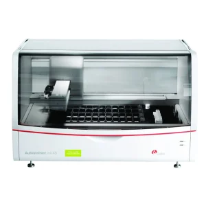
Autostainer Link Solution
Autostainer Link 48 provides labs with software and connectivity options that will greatly improve workload management and report generation while maintaining optimal immunohistochemistry staining results.

Hyperchrome
HyperChrome is the perfect Instrument for denaturation & hybridization procedures during FISH experiments.

VP2000
VP2000 is a consolidated workstation for
automated front-end FISH* processing
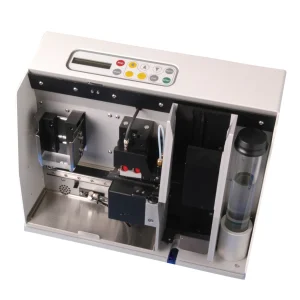
Dako Coverslipper
The Dako Coverslipper is a compact unit. It is small enough to fit inside fume cabinets and it can handle up to 600 slides per hour making it one of the fastest glass coverslippers currently available and setting a new benchmark in instrument design.
Testimonials & Reviews
Dr. (Prof.), Nitesh Mohan
Professor & Head, Department of Pathology, RMCH Bareilly
DSS's expertise, dedication, and professionalism were outstanding in making the Karyotyping & FISH workshop a great success. Their knowledge and valuable insights empowered all the participants with practical skills, receiving highly positive feedback from both students as well as faculty members.
Dr. Chhaya Chande, Professor & HOD, Microbiology
GGMCJJ Hospitals, Mumbai
“Ms. Megha Dhumal (Assistant Manager- Application) has done a satisfactory demonstration of the running of the Abbott Sample preparation machine model m2000sp and the Abbott RT-PCR machine model m2000rt. We appreciate the effort made by the DSS team under these difficult conditions to help our lab to carry out the imperative Covid-19 tests.”
Dr Sunil K Arora, Professor, Deptt of Immunopathology
PGIMER, Chandigarh
“We are using Confocal Microscope and one Fluorescence Microscope. Both are working fine. The after sales services by DSS have been excellent for functioning & upkeep of the microscopes. The applications support by experts from DSS is very useful. Keep it up!”
Dr Pramod Kumar Bajaj
MD, Spermprocessor Pvt Ltd
“Really excited to see the DSS Pathology solutions exhibition booth at APCON 2019 along with Magnus. We think all the upcoming technology had been displayed along with your efforts to make it Indigenous (Made in India) is highly appreciated. Wish you all the best. Keep it up!”
Dr. Sreejesh S, Associate Professor, Dept of Hematology
PGIMER, Chandigarh
“My experience with DSS so far has been very good till now. We are getting good support in both purchase as well as in troubleshooting. Very good experience with Mr Arun, Mr Manoj, Mr Mahesh and all others from the DSS team.”
Dr Sudha S Murthy, Department of Pathology and Laboratory Medicine
BIACH & RI, Hyderabad
“I am happy with DSS and associated with 19 years and use Dako antibody. Happy with Supply but need improvement.”
Dr S Radhika MD, PhD
Professor, Deptt. Of Cytology & Gynaec Pathology, PGIMER, Chandigarh
“PGI Cytology Dept. has had a long association with DSS- Olympus Microscopy Division. They have provided excellent services- after sales service. The product is also of very good quality. We have had no problems with their products and services are of very good quality.”
Dr Nuzhat Husain
RMLIMS, Lucknow
“Have been using Dako Reagents and Dako antibodies for a while. Services and products have been good and timely.”
Dr Minu Singh
Assistant Professor, PGIMER, Chandigarh
“MRC Holland MLPA products provided by DSS are of good quality, have never faced any quality issues with their product or shipping condition. They provide prompt response upon any query.”
Mr. Krishnani Professor, SGPGI, Lucknow
“My experience with DSS so far has been excellent for the last 30 years- sales and service experience. Microscope products are very useful and sturdy with high precision.”