DSS: Redefining Biotechnology & Life Science in India
- About Us
- Products & Services
PRODUCTS & SERVICES
-
Kits Reagents & Consumables
- Cytogenetics
- Dyes
- Fluorescence In Situ Hybridization (FISH)
- High-Performance Liquid Chromatography (HPLC)
- Histology
- Immuno Histo Chemistry (IHC)
- IVF Consumables
- Molecular Pathology & Diagnostics
- Multiplex Ligation-Dependent Probe Amplification (MLPA)
- Nucleic Acid Extraction
- PharmDx
- Real Time PCR
- Special Stains
- Instruments
- Software
- Accessories
- Advanced Material
-
Kits Reagents & Consumables
- Applications & Specialities
All Applications & Specialities
- Brands
- Contact Us
-

-
 0
0
- ☰
- About Us
- Products & Services
-
Kits Reagents & Consumables
- Cytogenetics
- Dyes
- Fluorescence In Situ Hybridization (FISH)
- High-Performance Liquid Chromatography (HPLC)
- Histology
- Immuno Histo Chemistry (IHC)
- IVF Consumables
- Molecular Pathology & Diagnostics
- Multiplex Ligation-Dependent Probe Amplification (MLPA)
- Nucleic Acid Extraction
- PharmDx
- Real Time PCR
- Special Stains
- Instruments
- Software
- Accessories
- Advanced Material
-
Kits Reagents & Consumables
- Applications & Specialities
- Brands
- Brand - Life Sciences
- 3i
- ABBERIOR INSTRUMENTS
- Abbott Molecular
- ADS Biotec
- APPLIED SPECTRAL IMAGING
- BioAir Tecnilabo
- DAKO (AGILENT)
- Eden Tech
- Elveflow
- ENTROGEN
- EUROCLONE
- EVIDENT
- Genea
- Hamamatsu Photonics
- Invivoscribe
- MASTER DIAGNOSTICA
- MBF BIOSCIENCE
- Medical Tek Co. Ltd
- MILESTONE MED SRL
- Molecular Machines & Industries
- MRC HOLLAND
- NeoDx
- Onward Assist
- Profound
- SCIENTIFICA
- SpaceGen
- Seqlo
- µCyte
- Brand - Industrial
- Brand - Life Sciences
- News & Events
- Career
- Contact Us
- Testimonial
- Blogs
- R&D
- CSR
- Press Release

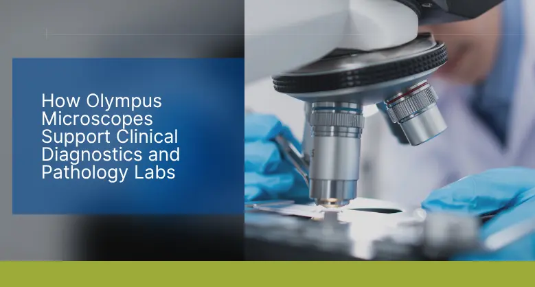
How Olympus Microscopes Support Clinical Diagnostics and Pathology Labs
BY DSS Imagetech Pvt Ltd 26th November 2025
In the world of modern medicine, the clinical diagnostics laboratory is the engine room of the healthcare system. It is a high-stakes, high-pressure environment that operates largely unseen by the patient, yet it is where the vast majority of medical decisions are born. Every day, this hub receives a constant stream of human samples, blood, tissue, and fluid, each one carrying an urgent, unanswered question. Is it an infection? Is it an autoimmune disease? Is it cancer? The answers provided by the laboratory will determine the patient’s prognosis and chart the course of their treatment.
At the very heart of this complex world, standing at the critical junction between the sample and the diagnosis, is the microscope. For the pathologist, the cytotechnologist, and the medical lab scientist, the microscope is not just a tool; it is a direct extension of their senses, their primary window into the patient’s biology. The quality, clarity, and reliability of the image they see are paramount. There is no room for ambiguity; there is no tolerance for optical distortion. A definitive diagnosis depends entirely on the faithful representation of the cells on the slide.
This is the world for which Olympus Microscopes were built. For over a century, the Olympus name has been synonymous with precision optics, engineering, and reliability. In the demanding ecosystem of the Olympus microscope pathology lab, these instruments are not just equipment; they are trusted partners in a daily quest for answers. From routine screening to the most complex molecular analysis, the Olympus portfolio offers a comprehensive and scalable solution designed to support every facet of the diagnostic workflow.
The microscope is the central tool in the diagnostic workflow
Before we can explore the specific hardware, we must first appreciate the diversity of tasks a microscope must perform in an Olympus microscope clinical diagnostics setting. The modern lab is not a single entity but a collection of specialized departments, each with unique needs.
In haematology, a technologist scans a blood smear, meticulously identifying and counting different types of white blood cells. They are looking for subtle changes in cell morphology, the shape of the nucleus, the color of the granules—that could signal a parasitic infection, an allergic reaction, or a life-threatening leukemia.
In cytology, a screener examines a Pap smear, searching for the faint, dysplastic changes in cervical cells that signal a pre-cancerous condition. The clarity of the nuclear chromatin is everything.
In surgical pathology, a pathologist examines a wafer-thin slice of a tumor, stained with hematoxylin and eosin (H&E). They are in a race against time, often performing a “frozen section” analysis while the patient is still on the operating table. The surgeon is waiting for the answer to one question: “Is it benign or malignant?” The pathologist must distinguish a normal cell from a cancerous one, a decision that will determine if the surgeon’s next step is to close the incision or perform a life-altering radical resection.
In all these scenarios, the microscope is the conduit for critical information. It must provide a perfectly flat, brilliantly illuminated, and colour-true image. It must be comfortable enough to be used for hours on end without causing fatigue. And it must be reliable enough to function flawlessly, day after day, year after year. This is the baseline expectation that defines the diagnostic environment.
Olympus upright microscopes are the daily workhorses for slide analysis
The most common sight in any clinical or pathology lab is the upright light microscope. This is the instrument that forms the backbone of daily testing, and the Olympus BX and CX series are industry standards.
The CX series, including the Olympus cx23 and Olympus cx43, represents the frontline of diagnostic work. These microscopes are designed for reliability, ease of use, and outstanding optical quality in a compact, efficient frame. The Olympus cx23 is a powerhouse of routine microscopy and education. It is frequently found in training labs for medical students and technologists, as well as in smaller clinics for daily urinalysis or blood smear analysis. Its robust build, long-life LED illumination, and field-number 20 objectives provide bright, clear images across a wide field of view, making screening fast and comfortable.
The Olympus cx43 steps up from this, offering a highly versatile and ergonomic platform for more demanding clinical environments. It is a popular choice for haematology and cytology labs. Its advanced color correction and uniform illumination are critical for differentiating the subtle shades of purple, pink, and blue in Giemsa- or PAP-stained slides. Its low-positioned stage and focus knobs reduce arm and shoulder strain, a vital feature for technicians who process hundreds of slides per day.
For the high-volume, high-complexity world of the Olympus microscope pathology lab, the BX series is the definitive workhorse. Models like the Olympus bx41, Olympus bx43, and Olympus bx53 are built on a platform of unmatched optical excellence and modularity. The Olympus bx41 and its successor, the Olympus bx43, are renowned for their stability and superior plan-apochromat objectives. These objectives ensure that the entire field of view, from the centre to the very edge, is perfectly in focus and free from color distortion. When a pathologist is scanning a slide for a small, isolated cluster of metastatic cells, this “flat field” is not a luxury; it is a necessity.
The Olympus microscopes has been a long-standing benchmark for advanced clinical analysis. Its modular frame allows it to be configured for a vast range of applications, from simple brightfield to complex, multi-head teaching systems and advanced fluorescence. Its ability to produce consistently brilliant, high-resolution images has made it a favourite in research-heavy diagnostic labs. Its evolution, the Olympuss bx53, takes this modularity even further. The Olympus bx53 is a truly state-of-the-art platform, often equipped with motorised components (like a motorized nosepiece or stage) and advanced LED illumination that provides true-to-life color rendering. This is critical in pathology, where the exact shade of an H&E or special stain provides vital diagnostic clues.
The Olympus BX46 was designed specifically to address the physical demands of pathology
Perhaps no other microscope demonstrates a deeper understanding of the pathology workflow than the Olympus BX46. The keyword that defines this instrument is ergonomics.
To understand why the Olympus BX46 microscope pathology combination is so specific, one must first humanise the problem. A pathologist does not just use a microscope; they inhabit it. They spend the majority of their professional life—often six to eight hours a day—hunched over, peering through eyepieces. This sustained, unnatural posture is a primary cause of chronic, work-related musculoskeletal disorders, with severe pain in the neck, back, and shoulders being a common complaint.
A fatigued pathologist is less effective than a well-rested one. Physical discomfort can break concentration, slow down throughput, and ultimately compromise diagnostic accuracy.
The Olympus BX46 was engineered from the ground up to solve this problem. Unlike a conventional microscope, where the user must adjust their body to the instrument, the BX46 is designed to adjust to the user’s body. It breaks from traditional design in several key ways:
- A Tilting, Telescoping Observation Tube: This allows the user to set the eyepiece height and angle perfectly, whether they are tall or short, eliminating the need to hunch.
- Low-Positioned Focus Knobs: The focus controls are placed low and close to the user, allowing their hands to rest comfortably on the desk, not “float” in the air.
- A Fixed, Low Stage: In a truly innovative move, the BX46 features a fixed-position stage. To focus, the user turns the knob, which moves the objective nosepiece up and down, not the stage. This means the stage—and the patient’s slide—always remains at the same comfortable, low height.
This focus on ergonomics is not a “comfort” feature. It is a core performance and safety feature. By dramatically reducing physical strain, the Olympus BX46 allows pathologists to work with sustained focus and comfort, directly supporting diagnostic confidence and lab productivity.
Olympus stereo microscopes are essential for 3D specimen analysis before a slide is even made
The diagnostic journey does not begin with a glass slide. It begins with the patient’s specimen, which is often a large, complex piece of tissue from a surgical resection. Before a 5-micron-thick slice can be cut for a slide, a pathologist or pathologist’s assistant must first perform “grossing,” or a gross examination.
This is where the Olympus stereo microscope becomes the essential tool. A stereo microscope provides a three-dimensional, lower-magnification view, which is exactly what is needed to examine a large specimen. The pathologist uses an instrument like the Olympus szx7 to look for the tumor, measure its size, assess how close it is to the surgical margins, and—most importantly—select the specific, representative sections that will be processed into slides.
The Olympus szx7 is a common sight at grossing stations because it provides a large zoom range and a wide field of view, with excellent depth of field. This allows the user to see the complex, 3D architecture of the tissue. Is the tumor invading the surrounding fat? Has it broken through the organ’s capsule? The answers to these questions, found with an Olympus stereo microscope, are critical for cancer staging and are just as important as the information found on the slide itself.
The Olympus fluorescence microscope opens a new world of molecular diagnostics
While the H&E stain is the workhorse of pathology, it cannot answer every question. Modern diagnostics increasingly rely on identifying specific proteins, genetic markers, and molecular pathways. This is the domain of the Olympus fluorescence microscope.
Fluorescence microscopy is a powerful technique that uses high-energy light to excite fluorescent probes (or “fluorophores”) that have been attached to specific cellular targets. The microscope’s filter system then allows only the emitted light from that probe to reach the user’s eye, resulting in a bright, glowing signal against a pitch-black background.
This has direct clinical applications:
- Immunofluorescence (IF): In nephrology (kidney diagnostics), a pathologist will use an Olympus fluorescence microscope (often a configured Olympus bx53) to look at a kidney biopsy. The glowing patterns of antibody deposits (IgA, IgG, C3) create a “fingerprint” that allows for the precise diagnosis of different types of glomerulonephritis.
- Fluorescence In Situ Hybridisation (FISH): In cancer diagnostics, FISH is used to find specific genetic abnormalities. For example, in breast cancer, it can determine if a tumor has an amplification of the HER2 gene. A positive result (seen as multiple bright signals in the cell nucleus) makes the patient eligible for a life-saving targeted therapy.
This level of analysis requires exceptional optical quality. The microscope must have objectives with high numerical apertures to gather as much faint light as possible, and the filter cubes must be perfectly optimized to separate the excitation and emission signals. Olympus‘s high-end uprights are engineered to excel at this, providing the high-contrast, low-noise images that molecular diagnostics demand.
This same need for specialized optics extends to the Olympus inverted microscope. While less common in a typical pathology lab, the inverted microscope is the standard in clinical cell culture labs (e.g., for cytogenetics or virology) and in fertility clinics. Its design—with the objectives below the stage, allows the user to look up at living cells in the bottom of a large culture flask or petri dish, which is impossible with a standard upright scope.
The Olympus digital microscope is transforming lab collaboration and analysis
The final piece of the modern lab is connectivity. The microscope is no longer a standalone, analogue island. The Olympus digital microscope represents the fusion of world-class optics with powerful digital imaging and software.
This is not a single model but an integrated system. A high-resolution Olympus digital camera attached to an Olympus bx43 or bx53 transforms the instrument from a personal viewing device into a powerful data-sharing hub. This capability is fundamentally changing the Olympus microscope clinical diagnostics workflow.
- Telepathology and Consultation: A pathologist at a regional hospital can capture a high-definition image of a difficult case and securely send it to a sub-specialist at a major academic center for a second opinion. This provides patients with access to world-class expertise, regardless of their location.
- Tumor Boards: Instead of all doctors huddling around a multi-head microscope, a pathologist can project a live, crystal-clear image of a slide onto a large screen for a multidisciplinary tumor board. This allows surgeons, oncologists, and radiologists to discuss the case and formulate a consensus treatment plan while all looking at the same key diagnostic features.
- Documentation and Reporting: High-quality images can be archived and embedded directly into a patient’s pathology report, providing a clear, permanent visual record of the diagnosis.
- Quantitative Analysis: This is the leap into computational pathology. Digital software can now perform objective analysis on an image. For example, in breast cancer, it can analyze an immunohistochemistry (IHC) slide and provide an exact percentage of cells that are positive for estrogen receptors (ER). This “H-score” is a critical data point that determines a patient’s hormonal therapy. This moves pathology from a qualitative art to a quantitative science, improving standardization and accuracy.
Olympus microscopes remain a vital partner in the evolution of diagnostic medicine
From the macro 3D view of an Olympus stereo microscope at the grossing station to the routine efficiency of an Olympus cx43, the ergonomic precision of an Olympus BX46, and the molecular insights of an Olympus fluorescence microscope, the Olympus portfolio provides a comprehensive, end-to-end ecosystem. These instruments are not just passive viewers; they are active partners in the diagnostic process.
They support the Olympus microscope pathology lab by delivering uncompromising image quality, which is the bedrock of diagnostic confidence. They support the lab’s personnel by providing ergonomic, fatigue-reducing designs that promote sustainable and accurate work. And they support the lab’s future by integrating seamlessly into a digital workflow, paving the way for telepathology, artificial intelligence, and a more collaborative, data-driven approach to medicine.
Behind every Olympus Microscope in a clinical lab, there is a patient anxiously awaiting an answer. The unwavering reliability and optical clarity of these instruments are a vital link in the chain of care, providing the answers that physicians need and the hope that patients deserve.
FAQs :-
1. How do Olympus Microscopes support clinical diagnostics and pathology labs?
Olympus Microscopes play a central role in clinical diagnostics by providing unmatched optical clarity, reliability, and precision. They assist pathologists and lab technologists in accurately identifying infections, autoimmune diseases, and cancers. With models designed for every stage of diagnostics —from sample screening to molecular analysis—Olympus ensures confident and timely diagnoses.
2. What are the key microscope models used in pathology and their specific applications?
The Olympus CX series (CX23, CX43) is ideal for routine lab work and education due to its ease of use and reliable optics. The BX series (BX41, BX43, BX53) is designed for advanced diagnostics and research, offering superior image quality and modular flexibility. The BX46 focuses on ergonomic design to reduce fatigue during long hours of slide observation.
3. Why is ergonomics important in pathology microscopy, and how does the Olympus BX46 address it?
Pathologists spend long hours using microscopes, leading to neck, back, and shoulder strain. The Olympus BX46 is engineered to minimize physical fatigue with features like a tilting telescoping observation tube, low-positioned focus knobs, and a fixed low stage. These ergonomic innovations enhance comfort and support sustained focus for accurate diagnostics.
4. How are Olympus stereo and fluorescence microscopes used in diagnostics?
The Olympus SZX7 stereo microscope is essential for 3D specimen analysis during gross examination, helping pathologists identify and sample key tissue areas. The Olympus fluorescence microscopes (e.g. BX53) are vital for molecular diagnostics, enabling techniques like Immunofluorescence (IF) and Fluorescence In Situ Hybridization (FISH) to detect specific proteins and genetic abnormalities in diseases such as kidney disorders and cancer.
5. How are digital Olympus microscopes transforming modern pathology labs?
Digital integration allows Olympus microscopes to capture, share, and analyze high-resolution images for telepathology, tumor boards, and quantitative analysis. They enable real-time collaboration, digital record-keeping, and AI-assisted image quantification, transforming traditional microscopy into a connected, data-driven diagnostic workflow.
Latest Articles
World AIDS Day: Breaking the Stigma and Understanding HIV Testing
BY DSS Imagetech Pvt Ltd December 1, 2025
Worlds AIDS Day 2025 focuses on the theme “Overcoming disruption, transforming the AIDS response.” Highlighting the need for a stronger, more resilient approach to end the epidemic. This theme acknowledges...
Read MoreHow Olympus Microscopes Support Clinical Diagnostics and Pathology Labs
BY DSS Imagetech Pvt Ltd November 26, 2025
In the world of modern medicine, the clinical diagnostics laboratory is the engine room of the healthcare system. It is a high-stakes, high-pressure environment that operates largely unseen by the...
Read MoreThe Role of Genetic Testing (BRCA, Onco-panels) in Breast Cancer...
BY DSS Imagetech Pvt Ltd November 18, 2025
Breast cancer is a complex and deeply personal diagnosis that will affect many women in their lifetime. For decades, our primary approach was reactive: focusing on awareness, monthly self exams,...
Read More


