DSS: Redefining Biotechnology & Life Science in India
- About Us
- Products & Services
PRODUCTS & SERVICES
-
Kits Reagents & Consumables
- Cytogenetics
- Dyes
- Fluorescence In Situ Hybridization (FISH)
- High-Performance Liquid Chromatography (HPLC)
- Histology
- Immuno Histo Chemistry (IHC)
- IVF Consumables
- Molecular Pathology & Diagnostics
- Multiplex Ligation-Dependent Probe Amplification (MLPA)
- Nucleic Acid Extraction
- PharmDx
- Real Time PCR
- Special Stains
- Instruments
- Software
- Accessories
- Advanced Material
-
Kits Reagents & Consumables
- Applications & Specialities
All Applications & Specialities
- Brands
- Contact Us
-

-
 0
0
- ☰
- About Us
- Products & Services
-
Kits Reagents & Consumables
- Cytogenetics
- Dyes
- Fluorescence In Situ Hybridization (FISH)
- High-Performance Liquid Chromatography (HPLC)
- Histology
- Immuno Histo Chemistry (IHC)
- IVF Consumables
- Molecular Pathology & Diagnostics
- Multiplex Ligation-Dependent Probe Amplification (MLPA)
- Nucleic Acid Extraction
- PharmDx
- Real Time PCR
- Special Stains
- Instruments
- Software
- Accessories
- Advanced Material
-
Kits Reagents & Consumables
- Applications & Specialities
- Brands
- Brand - Life Sciences
- 3i
- ABBERIOR INSTRUMENTS
- Abbott Molecular
- ADS Biotec
- APPLIED SPECTRAL IMAGING
- BioAir Tecnilabo
- DAKO (AGILENT)
- Eden Tech
- Elveflow
- ENTROGEN
- EUROCLONE
- EVIDENT
- Genea
- Hamamatsu Photonics
- Invivoscribe
- MASTER DIAGNOSTICA
- MBF BIOSCIENCE
- Medical Tek Co. Ltd
- MILESTONE MED SRL
- Molecular Machines & Industries
- MRC HOLLAND
- NeoDx
- Onward Assist
- Profound
- SCIENTIFICA
- SpaceGen
- Seqlo
- µCyte
- Brand - Industrial
- Brand - Life Sciences
- News & Events
- Career
- Contact Us
- Testimonial
- Blogs
- R&D
- CSR
- Press Release
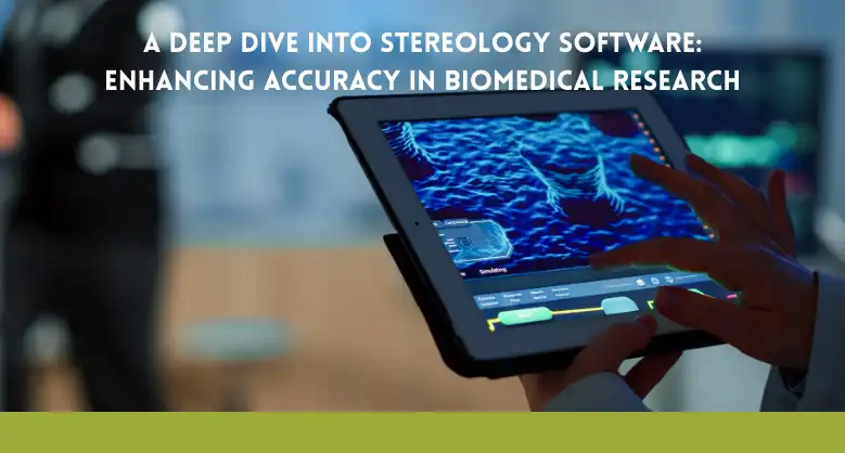
A Deep Dive into Stereology Software: Enhancing Accuracy in Biomedical Research
BY admin 29th August 2025
Have you ever considered how many scientific breakthroughs hinge on a seemingly simple question: “How many?” From counting neurons in a mouse’s brain to measuring the volume of a tumor, the numbers we collect in biomedical research form the foundation of our understanding. But what if those numbers were wrong? What if the methods we rely on to get these vital counts and measurements were flawed, introducing hidden biases that skewed our results? This isn’t just a hypothetical concern; it’s a real and often overlooked challenge in laboratories worldwide. In this post, we’ll dive into the world of stereology software, exploring how it’s revolutionizing the way we get answers to these crucial questions, and we’ll uncover some surprising truths about what it takes to get truly accurate data.
The Problem with Traditional Counting: Why Your Data Might Be Flawed
For years, many researchers relied on simple, model-based methods to count cells or measure structures in tissue samples. These methods often involved counting everything in a single slice or making assumptions about the objects being studied’s size, shape, and distribution. While seemingly straightforward, these approaches are fraught with potential for error. Imagine counting the number of trees in a forest by only looking at a single, thin line drawn through it. You’d likely miss many trees and overcount others that are only partially on the line. The same problem happens when looking at a thin tissue slice under a microscope.
The core issue is that our 2D observations don’t always accurately represent the 3D world. You can count a cell’s profile multiple times as it appears in different slices, or you may miss smaller cells. The human eye, too, introduces its form of bias; we tend to prefer looking at what’s easy to see, which can lead to counting more of the larger, more prominent structures. These hidden assumptions, or “model-based” biases, can lead to inaccurate and unreliable results that are difficult to reproduce and may even lead to incorrect scientific conclusions.
Enter Design-Based Stereology: The Science of Unbiased Sampling
To address these fundamental problems, a new way of thinking emerged: design-based stereology. This robust set of mathematical principles and tools allows scientists to get accurate, unbiased, and reproducible data without making assumptions about the size, shape, or distribution of their study objects.
Think of it like this: instead of trying to count every single tree in the forest, you would use a carefully designed, systematic random sampling method. You may split the woodland into a grid and randomly choose a few areas to determine the trees therein. Following a strict set of rules and using a specific geometric “probe” (like a counting frame or a grid of points), you can then use a formula to get a statistically accurate prediction of the forest’s total tree count.
Design-based stereology applies this same logic to the microscopic world. It uses a series of carefully planned steps to ensure that every object in your sample—a cell, a blood vessel, or a tumor—has an equal chance of being selected and counted. This eliminates the biases of traditional counting methods and gives you results you can trust.
Stereology software has the ability to simplify complex methods.
While the principles of stereology are sound, performing these calculations by hand would be incredibly tedious and time-consuming. This is where stereology software comes in. It’s the essential tool that brings the power of design-based stereology to the modern lab.
These specialized software systems are designed to guide the user through the entire process, from setting up the experiment to collecting the data and performing the final calculations. They automate the most difficult parts of stereology, such as the systematic random sampling of the tissue and the complex mathematical calculations. This saves a tremendous amount of time and minimizes the chance of human error.
A great example of this is the Stereo Investigator software system. It’s a comprehensive platform integrates with your microscope, controlling the motorized stage and camera to perform systematic random sampling automatically. It provides a user-friendly interface with different “probes” for various types of measurements, such as:
- Optical Fractionator Probe: This is the gold standard for getting an unbiased count of the total number of cells in a region.
- Cavalieri Estimator: Used to get an unbiased estimate of the volume of a structure, like a brain region or a tumor.
- Spaceballs Probe: A clever tool for measuring the length of thin, winding structures like blood vessels or nerve fibers.
Using these specialized tools, researchers can get precise estimates of crucial parameters like cell counts, volumes, and lengths, all with a built-in measure of accuracy called the coefficient of error (CE). The software calculates this for you, so you always know how reliable your data is.
Who Uses Stereology Software and Why It Matters
The Global Reach of Stereology: A Look at MBF Bioscience India
The demand for accurate research tools is not limited to any single country. The need for reliable, unbiased data is a global constant, especially in a rapidly growing scientific hub like India. As biomedical research and pharmaceutical development flourish in the country, so does the need for advanced tools like stereology software.
Companies like MBF Bioscience India are crucial in meeting this demand. They provide researchers with access to robust systems like Stereo Investigator and the support and expertise needed to implement these complex methods correctly. This local presence is vital because scientists can get training, technical support, and guidance in their region and language.
The availability of these tools in India is a game-changer for local research. It allows scientists to conduct studies that meet the highest international standards, publish in top-tier journals, and contribute to the global body of scientific knowledge with beyond reproach data. It ensures that the biomedical research coming out of India is as accurate and impactful as any in the world.
The Future is Clear: Combining Stereology with New Technologies
As technology continues to advance, so does the potential of stereology. We are seeing exciting developments that combine the rock-solid principles of design-based stereology with modern imaging techniques.
One of the most significant recent advances is the ability to perform stereology on cleared tissue. In the past, tissue samples had to be sliced into thin sections for analysis, which could lead to damage and information loss. New clearing techniques make tissue transparent, allowing imaging of the entire 3D structure using tools like light sheet or confocal microscopy. This has opened up a new world of possibilities, enabling scientists to get unbiased data from intact tissue specimens. Stereo Investigator has adapted to this change, with a Cleared Tissue Edition explicitly designed for advanced imaging.
Another area of growth is the integration of stereology with artificial intelligence (AI) and machine learning. While AI can be a useful tool for swiftly recognizing and counting objects, it is not flawless and may carry biases. The future of research will likely see a hybrid approach, where AI is used for rapid, high-throughput screening, stereology is used to validate the most critical measurements, and training and refining the AI algorithms is done. This combination of speed and accuracy will be a powerful force in accelerating scientific discovery.
How to Choose the Right Stereology Software for Your Lab
If you’re a researcher looking to improve the accuracy of your quantitative data, choosing the right stereology software is a critical decision. Here are some key factors to consider:
- Integration with Your Existing Equipment: Does the software work with your current microscope, motorized stage, and camera? A sound system should be compatible with various hardware from different manufacturers.
- Ease of Use and Training: While stereology is a complex field, the software should make the process as straightforward as possible. Look for systems with intuitive workflows, clear guides, and robust training resources.
- Comprehensive Set of Probes: The software should offer a full suite of design-based probes for different types of measurements (e.g., cell counting, volume, length, surface area).
- Support and Community: Does the company provide excellent technical and scientific support? Is there a community of users you can learn from? Having access to expert guidance is invaluable, especially when you’re first getting started.
- Reputation and Citations: Look at how often the software is cited in peer-reviewed publications. A widely cited system like Stereo Investigator software is a good indicator of its reliability and acceptance within the scientific community.
By carefully considering these factors, you can find a solution that not only enhances the accuracy of your research but also streamlines your workflow and helps you produce publishable-quality data.
Conclusion: A New Standard for Accuracy in Biomedical Research
Ultimately, the question isn’t just “how many?” but “how do we know our numbers are right?” Stereology software provides the definitive answer to that question. By moving away from biased, model-based counting and embracing the rigorous, assumption-free principles of design-based stereology, researchers can generate more accurate, reproducible, and trustworthy data.
From the research labs of Europe to the growing scientific communities in places like MBF Bioscience India, these tools are setting a new standard for quantitative analysis. Integrating stereology with modern technologies like tissue clearing and AI promises to accelerate our understanding of disease further and open up new avenues for discovery. The next time you see a scientific paper with a graph of cell counts or a measurement of tissue volume, you can be sure that behind those numbers lies a commitment to accuracy, a commitment often made possible by the powerful, unbiased approach of stereology.
Faq’s :-
Q1. What is stereology software, and why is it important in biomedical research?
Stereology software is a specialized tool that helps researchers obtain accurate, unbiased, and reproducible data from tissue samples. Instead of relying on error-prone, model-based counting methods, design-based stereology ensures every cell or structure has an equal chance of being sampled. At DSS Imagetech, we provide advanced stereology solutions that support neuroscientists, oncologists, toxicologists, and stem cell researchers in generating reliable data for high-impact studies.
Q2. How does stereology software improve accuracy compared to traditional counting methods?
Traditional methods often rely on 2D slices and visual assumptions, which can lead to over- or under-counting. Stereology software eliminates these biases by using systematic random sampling and mathematical probes like the Optical Fractionator, Cavalieri Estimator, and Spaceballs Probe. DSS Imagetech offers stereology platforms that integrate seamlessly with microscopes, ensuring unbiased data for cell counts, tumor volumes, and structural measurements.
Q3. What biomedical research areas benefit most from stereology solutions provided by DSS Imagetech?
Stereology software is widely used across multiple disciplines: – Neuroscience: Counting neurons in Alzheimer’s and Parkinson’s studies. – Oncology: Measuring tumor volume and vascularization. – Toxicology: Assessing structural changes in organs after drug testing. – Pulmonary research: Quantifying alveoli and lung surface area. – Stem cell research: Tracking integration of transplanted cells. Through DSS Imagetech, Indian labs can access globally recognized stereology tools and expertise for these applications.
Q4. How does DSS Imagetech support researchers using stereology software in India?
DSS Imagetech partners with leading global brands like MBF Bioscience to deliver stereology software and hardware solutions in India. Beyond supplying tools like Stereo Investigator, our team provides installation, training, and scientific support. This ensures Indian researchers can conduct studies that meet international standards, publish in reputed journals, and contribute impactful findings backed by accurate quantitative analysis.
Q5. What should a lab consider before choosing stereology software from DSS Imagetech?
When selecting stereology solutions, researchers should look for: – Compatibility with samples, compataibility of existing microscopes and imaging systems – Ease of use with guided workflows and training resources. – Comprehensive probes for unbiased counts, volumes, lengths, and surface areas. – Technical support and expertise for smooth implementation. – Global reputation and citations in peerreviewed journals. DSS Imagetech ensures all these factors are met, helping laboratories adopt the right stereology platform for accurate and reproducible results.
Latest Articles
World AIDS Day: Breaking the Stigma and Understanding HIV Testing
BY DSS Imagetech Pvt Ltd December 1, 2025
Worlds AIDS Day 2025 focuses on the theme “Overcoming disruption, transforming the AIDS response.” Highlighting the need for a stronger, more resilient approach to end the epidemic. This theme acknowledges...
Read MoreHow Olympus Microscopes Support Clinical Diagnostics and Pathology Labs
BY DSS Imagetech Pvt Ltd November 26, 2025
In the world of modern medicine, the clinical diagnostics laboratory is the engine room of the healthcare system. It is a high-stakes, high-pressure environment that operates largely unseen by the...
Read MoreThe Role of Genetic Testing (BRCA, Onco-panels) in Breast Cancer...
BY DSS Imagetech Pvt Ltd November 18, 2025
Breast cancer is a complex and deeply personal diagnosis that will affect many women in their lifetime. For decades, our primary approach was reactive: focusing on awareness, monthly self exams,...
Read More