DSS: Redefining Biotechnology & Life Science in India


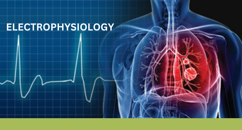
ELECTROPHYSIOLOGY
Electrophysiology studies the change in electrical properties of living neurons and investigates biological and molecular processes within the nervous system. In neuroscience communication between neurons is based on electrical and chemical signals. So, the electrophysiology technique records neuron activity and helps scientists to decode the intracellular and intercellular messages or any neuronal disorder by measuring the electrical activity of the cell.
Planar patch clamp is a novel electrophysiology tool developed for high throughput and to understand ion channel behaviour. It plays a key role in many neurological and cardiovascular diseases., as well as in physiological functions.
Electrophysiology lab
Every EP lab setup is different depending upon the requirement of the experiments where some equipment is mandatory and some are not.
An electrophysiological setup has four main lab requirements which are common in all the setups to record electrical signals from neurons.:
1. Environment — for keeping the preparation healthy;
2. Optics — visualizing the preparation;
3. Mechanics — stably positioning the microelectrode;
4. Electronics — amplifying and recording the signal.
Electrophysiology rig- It is an instrument used in electrophysiology techniques. The standard setup of the electrophysiology rig is shown here-
Microelectrode-Two types of electrodes are metal electrodes and glass micropipettes filled with an electrolyte solution. The signals are detected by these microelectrodes which further transfer signals to the amplifier, oscilloscope and computer.
Headstage-It is the central hub which connects the microelectrode with an amplifier to detect electrical signals and contains the electrode holder which stabilizes the recordings. The headstage is positioned with the help of a micromanipulator and attached to the Microdrive.
Micromanipulator-Allows movements in all the axes i.e., X, Y, and Z axes allowing the proper position of a microelectrode in tissue. A micromanipulator which has fine-scale units of measurement (usually μm) is good and can be used in specific regions of the brain or tissue
Microdrive-It is used to lower or raise microelectrode to a depth in very steps. Remote control Microdrive is used to reduce hand vibration. In very fine steps, the Microdrive lowers or raises the microelectrode at a specific depth in tissue.
The amplifier-The signal is then passed from the microelectrode on the headstage to the amplifier where amplification of the signal takes place. After that amplifier transfers signals to the oscilloscope or computer.
Oscilloscope-Receives signals from the amplifier and displays membrane potential over time. Any changes in electrical voltage can be heard on the loudspeaker.
Microscope-It is necessary for all kinds of physiological recordings. A low-power microscope is used to see morphological features of the brain and tissue. The inverted microscope is preferred over any other because it allows easy access to the sample and provides a larger platform for the micromanipulator.
Computer- The software in the computer makes it easy to write programs which can introduce electrical stimulus for tissue preparations and record neural responses, display results of the experiment even when an experiment is going on and also do real-time data analysis.

Electrophysiology Testing
EP testing is the most efficient parameter for the evaluation of nerves. It can be diagnostic, prognostic, and potentially therapeutic. This testing is primarily used in patients who have irregular heart rhythms (arrhythmia). supraventricular tachycardia (SVT) or any other type of tachycardia then EP study will provide the best treatment. Electrophysiology testing has some drawbacks like negative EP does not determine arrhythmia in a patient, and does not provide anatomic information about the peripheral nerve or location of lesion.
Faq’s :-
1. What is electrophysiology?
Electrophysiology is the study of electrical activity in biological cells and tissues, particularly in the heart and nervous system.
2. Why is electrophysiology important in medical science?
It helps doctors diagnose and treat conditions related to abnormal electrical activity in the body.
3. What diseases can electrophysiology help diagnose?
Electrophysiology is commonly used to diagnose heart rhythm disorders and neurological conditions.
4. What tests are used in electrophysiology studies?
Common tests include electrocardiograms (ECG), nerve conduction studies, and intracardiac electrophysiology testing.
5. How does electrophysiology help treat heart disorders?
It helps identify abnormal electrical pathways in the heart so doctors can treat arrhythmias effectively.
Latest Articles

World Tuberculosis Day: “Spreading Awareness, Saving Lives”
BY Mr. Satendra Saxena, Application Specialist, DSS Imagetech 23rd March 2026
Every time on March 24, the world pauses to observe World Tuberculosis (TB) Day. It’s a day of mixed feelings. We celebrate the scientific improvements that have made this complaint...
Read More
International Women’s Day – Prioritising Women’s Health for a Healthier...
BY Ms. Sohini Chatterjee, Application Specialist, DSS Imagetech 7th March 2026
In 2013, one public disclosure transformed the global conversation around women’s health. When Angelina Jolie revealed in a New York Times op-ed that she carried a BRCA1 mutation and had...
Read More
How BX51WI Microscopes Support High-Resolution Neural Imaging
BY DSS Imagetech Pvt Ltd 24th February 2026
Picture this scenario. It’s 6:00 PM on a Friday. You have spent the last six hours harvesting tissue. You perfused the mouse perfectly, the liver cleared instantly, and the brain...
Read More