DSS: Redefining Biotechnology & Life Science in India


Home > Microscopy & Imaging
Microscopy & Imaging
The Microscopy & Imaging division has been in existence since more than 40 years and is one of the foundation pillars of DSS. This long-standing experience in the business of Microscopy & Imaging has given the Microscopy & Imaging division a unique position as the most informed supplier of Microscopes and Digital Imaging solutions in India. As a complete solutions provider for microscope users across multiple user segments, DSS has developed unique skills and capabilities to configure and integrate Microscopy & Imaging based system solutions for life sciences research and clinical applications like Pathology IVF and Cytogenetics. To compliment the Microscopy & Imaging business DSS has successfully added to its portfolio a complete range of In-vivo imaging platforms.
The Microscopy & Imaging division ensures “Total Customer Satisfaction” by providing pan India support to our customers through our dedicated Application and Service teams.
The qualified and expert application team of Microscopy & Imaging is:
Dr. Anirban Bose has joined DSS Imagetech Pvt. Ltd. as a Product and Application Specialist for high-end imaging systems after completing his Ph.D. in Biochemistry from the University of Calcutta. His doctoral research concerns various biophysical techniques based on optical microscopy and spectroscopy and is conceded by a distinguished number of patents and publications in peer-reviewed journals. He also has expertise in developing autonomous optoelectronic sensing devices and their applications.
Ms. Nikita Kawli has done MSc (Applied Genetics), PGD MLT, PGD in Bio Nanotechnology She is working with DSS Imagetech as Product Application Manager covering a variety of Cytogenetics & Pathology Imaging products. Nikita has been involved in the entire cycle of Presales to post-sales activities. Professional competency in performing various assays on different samples, exposure to conducting various complicated tests to identify Infectious Diseases and Inheritable conditions, Calibration and Maintenance of various instruments, and Cytogenetics and Molecular Biological Works.
Dr. Shomnath Bhowmick is an expert in optics, material science, and advanced microscopy, with a strong focus on biological imaging and life sciences applications. Specializing in Confocal, STED, AFM, SEM, and TEM imaging, he has extensive experience in high-resolution characterization of biological and material systems. Holding a Ph.D. in Information Technology & Electrical Engineering (MEMS) from the University of Federico II, Naples, his research spans microfabrication, AFM-IR technique development, and bio-photonics. He has worked on MEMS resonators and thin-film characterization at SilTerra Malaysia and explored AFM, droplet microfluidics, and advanced imaging for biological applications at Shilps Sciences. Currently, as an Application Specialist & Sales Manager at DSS Imagetech, he provides technical expertise in AFM, Microfluidics, Confocal, Multiphoton, and STED microscopy, enabling cutting-edge research in cell biology, neuroscience, and bio-imaging. He holds multiple patents and has published extensively in leading scientific journals, contributing to advancements in biophysical imaging, nanoscale characterization, and photonics. His expertise extends to cleanroom processing, microelectronics, and computational modeling using COMSOL, MATLAB, and Python, bridging the gap between engineering, life sciences, and next-generation imaging technologies.
Dr. Sneha Paul completed her Ph.D. in Chemistry from University of Hyderabad, India. She is working with DSS Imagetech Pvt. Ltd. as an Application Specialist for high-end systems in the Micro Imaging Division. Prior to this, Sneha worked as a Postdoctoral Researcher at McGill University, Canada and University of Chicago, USA and as a Research Associate at CNRS-ISMO, France. She has expertise in advanced microscopic and spectroscopic techniques like confocal microscopy, super-resolution microscopy and transient absorption spectroscopy. She also has experience in developing image analysis algorithms.

Olympus BX63 Microscope
The BX63 offers outstanding stability and imaging precision for research applications, with the convenience of a touch screen interface for easy operation. Additionally, it features a detachable controller that can be positioned to suit the operator's preference or workflow requirements.
Applications & Specialities, Instruments, Microscopes, Microscopy, Products & Services, EVIDENT, Microscopy & Imaging
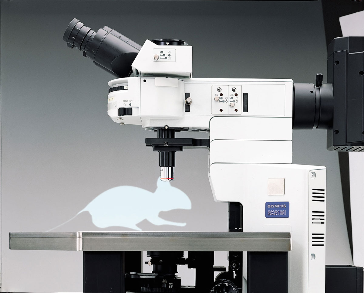
BX51WI
The BX51WI is a special-purpose research microscope designed for electrophysiology. The BX51WI provides the stability crucial for patch clamping and is equipped with dedicated water immersion objectives designed for near IR illumination at 775 and 990nm.
Applications & Specialities, Instruments, Microscopes, Microscopy, Products & Services, EVIDENT, Microscopy & Imaging
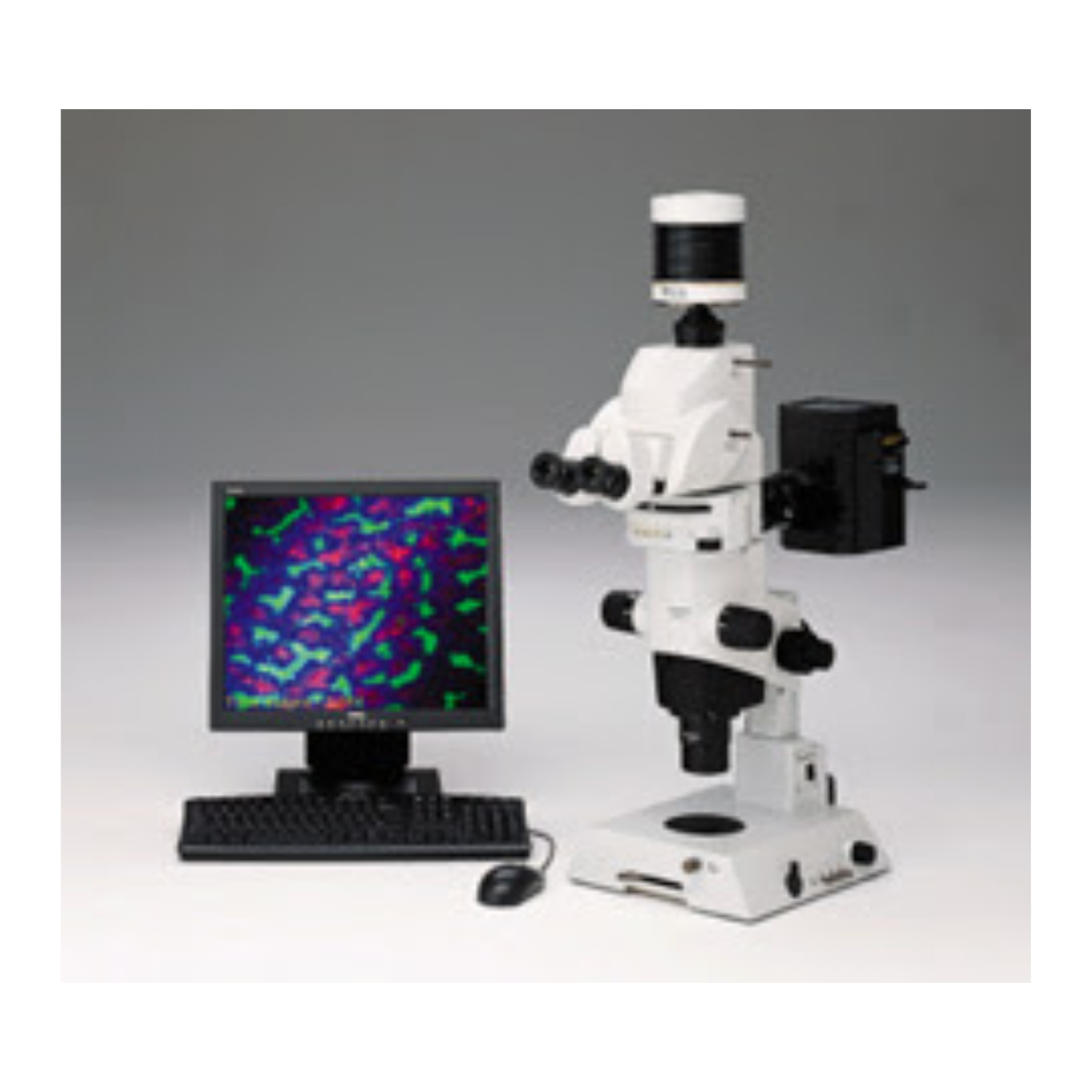
Olympus MVX10 Microscope
The MVX10 MacroView is a research Macro Zoom Fluorescence Microscope. The MVX10 addresses the tradeoffs between stereo and compound imaging systems, offering high numerical apertures from a compound microscope along with long working distances and larger fields of view from a stereo microscope.
Applications & Specialities, Instruments, Microscopes, Microscopy, Products & Services, EVIDENT, Microscopy & Imaging
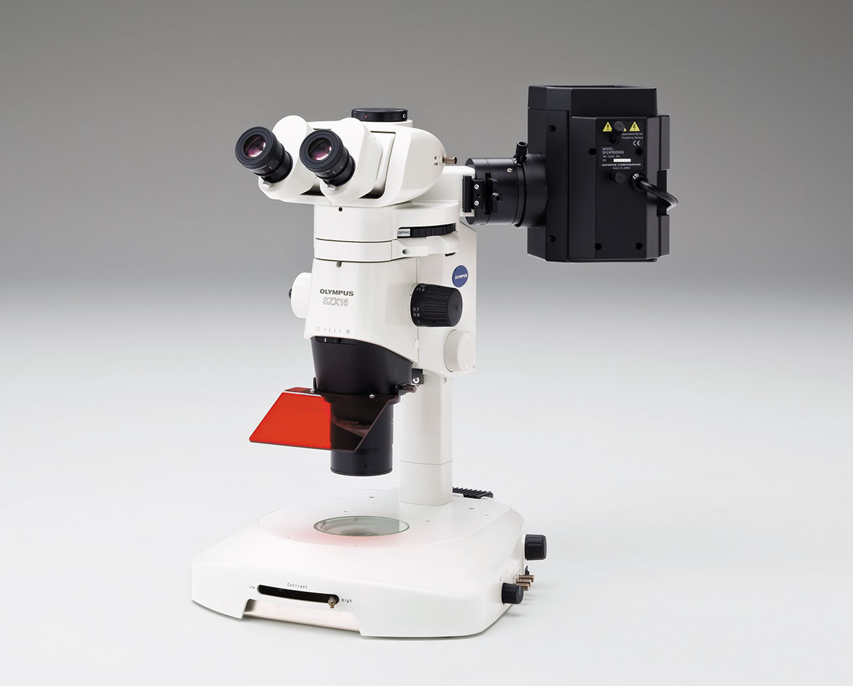
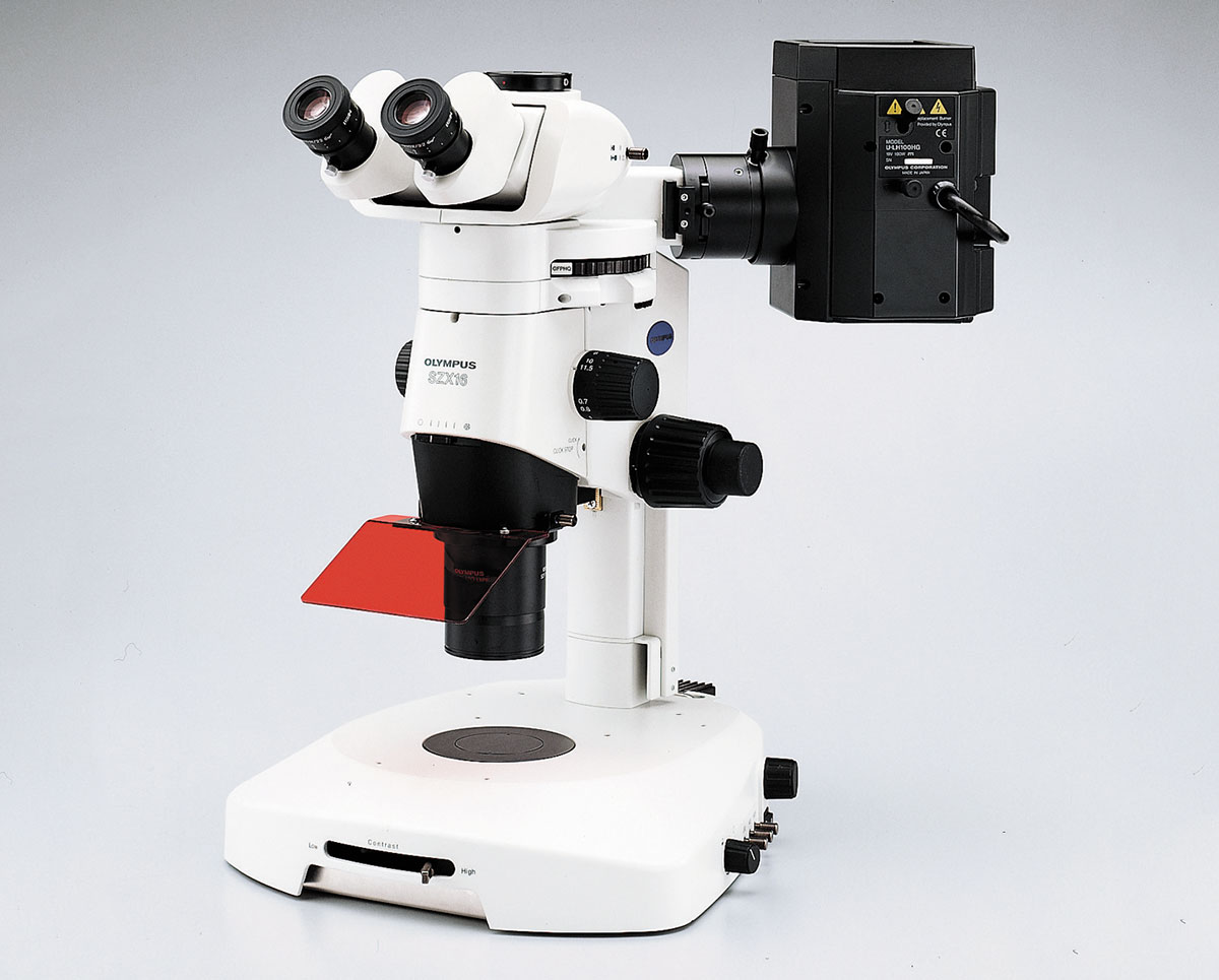
Olympus SZX16 Microscope
The new SZX16 is designed for advanced research and has a maximum numerical aperture of 0.30, producing a resolution of 900 line pairs per millimeter.
Applications & Specialities, Instruments, Microscopes, Microscopy, Products & Services, EVIDENT, Microscopy & Imaging
Olympus SZX10 Microscope
The Olympus SZX10 is designed for routine research. It offers a maximum numerical aperture (NA) of 0.2, producing a resolution of 600 line pairs per millimetre.
Applications & Specialities, Instruments, Microscopes, Microscopy, Products & Services, EVIDENT, Microscopy & Imaging
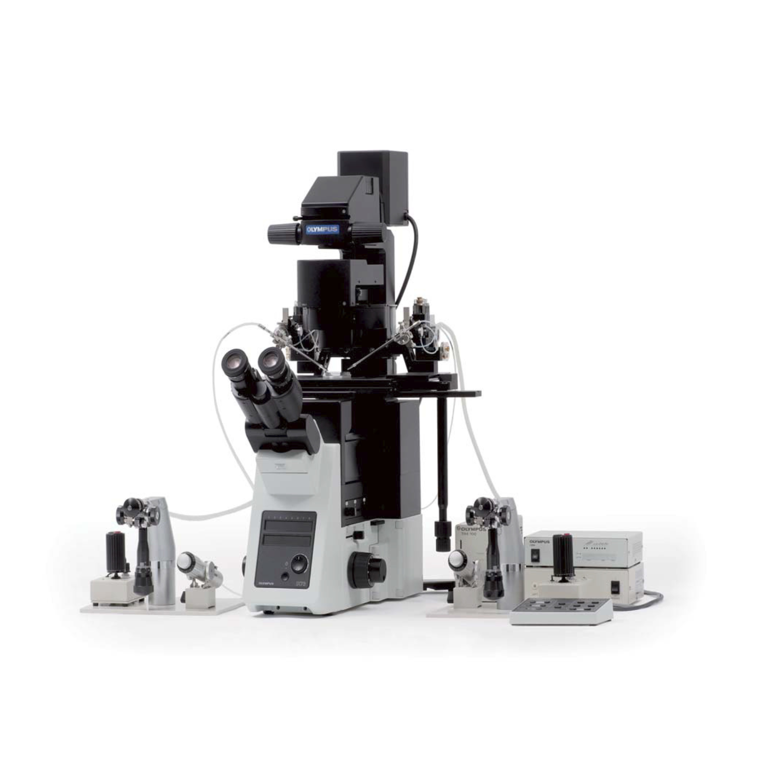
Olympus IX73 Inverted Microscope
The IX73 inverted microscope system sets new standards in advanced live cell imaging with its stable compact frame, outstanding optical performance and maximum flexibility.
Applications & Specialities, Instruments, Microscopes, Microscopy, Products & Services, EVIDENT, Microscopy & Imaging
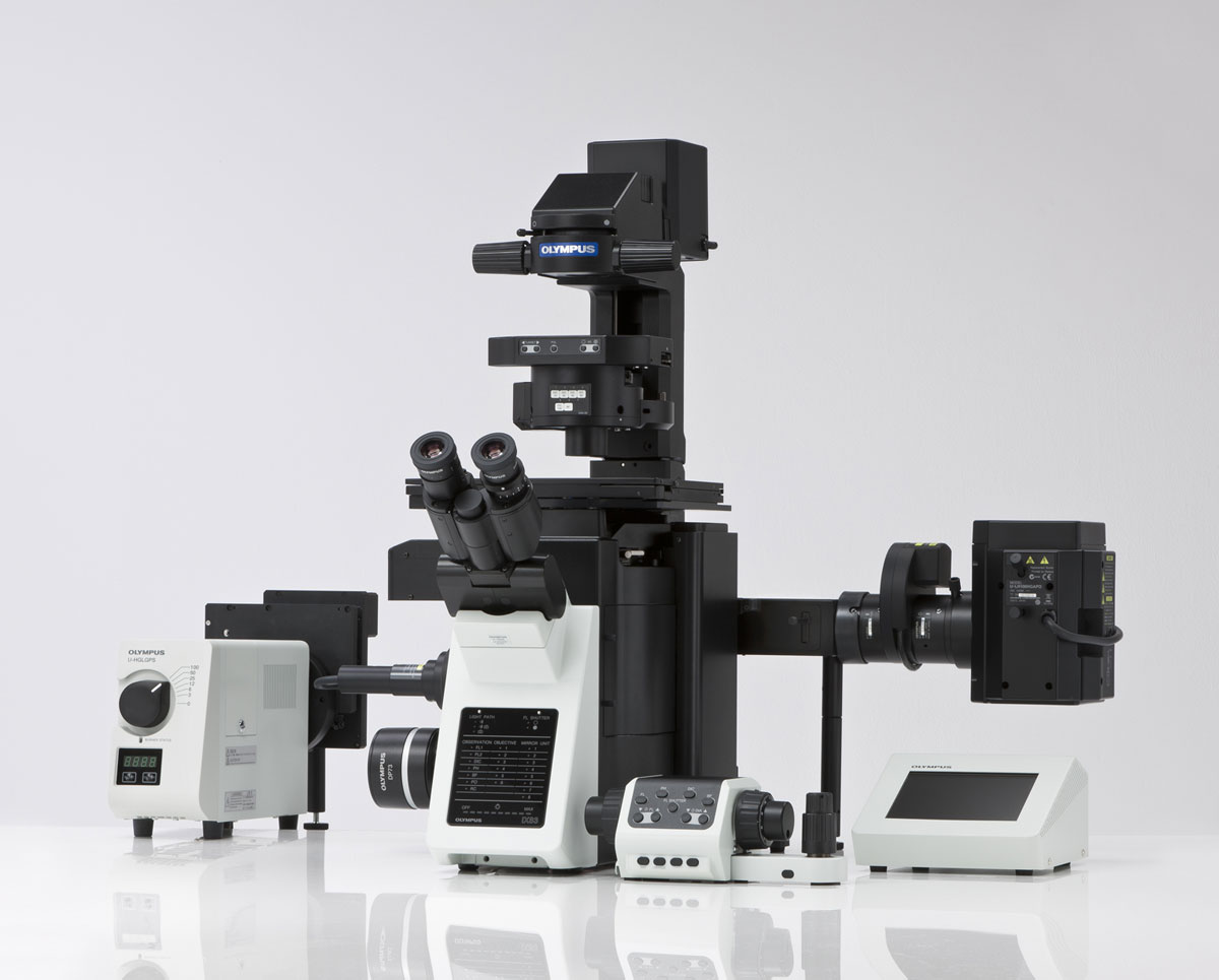
Olympus IX83 Microscope
The fully motorised and automated IX83 is the most advanced member of the IX3 series of imaging systems for life science researchers and offers a new level of usability and application flexibility.
Applications & Specialities, Instruments, Live Cell Imaging, Microscopes, Microscopy, Products & Services, EVIDENT, Microscopy & Imaging
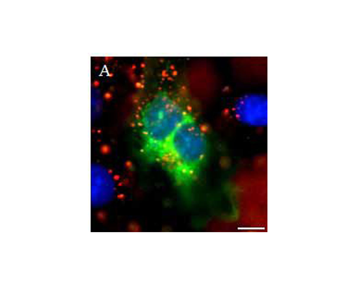
Spectraview
HyperSpectral imaging represents an enabling technology for a broad spectrum of applications in biomedical imaging.
Applications & Specialities, Oncology, Pathology, Products & Services, Software, APPLIED SPECTRAL IMAGING, Microscopy & Imaging
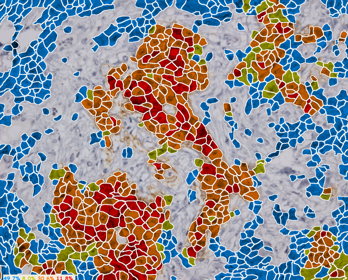
HiPath
GenASIs HiPath is a highly intelligent computerized scoring aid for quantitative immunohistochemistry and CISH assays that aides in review and analysis of tissue samples.
Applications & Specialities, Oncology, Pathology, Pathology Software, Products & Services, Software, APPLIED SPECTRAL IMAGING, Microscopy & Imaging
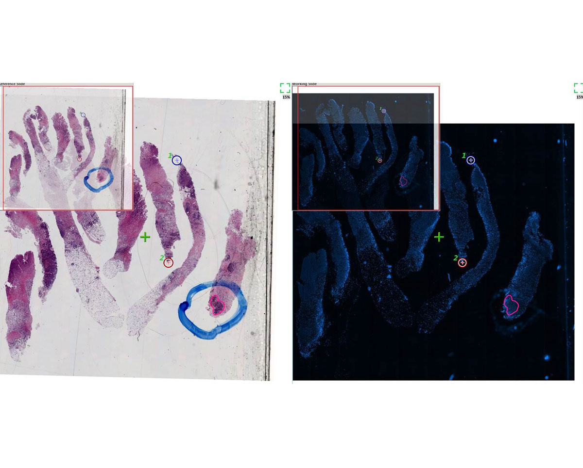
Tissue Match
Tissue Match is a tool for locating and marking a specific region of interest on FISH slides prior to scanning at high resolution.
Applications & Specialities, Oncology, Pathology, Products & Services, APPLIED SPECTRAL IMAGING, Microscopy & Imaging
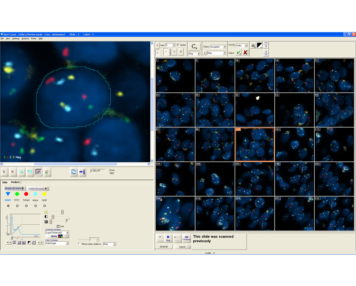
Spotscan (Solid FISH)
The GenASIs SpotScan application automatically analyzes FISH cells. While FISHView performs analysis on one cell at a time
Applications & Specialities, Oncology, Pathology, Pathology Software, Products & Services, Software, APPLIED SPECTRAL IMAGING, Microscopy & Imaging
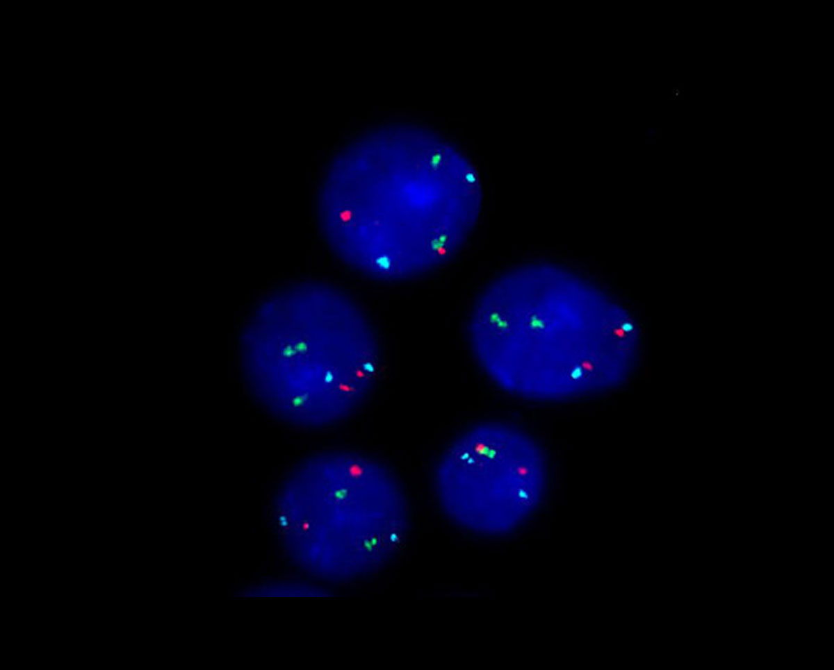
FISHview (Solid FISH)
GenASIs FISH is a powerful tool for FISH technologists working in pathology and cytogenetic labs. With GenASIs FISH, labs can work faster and provided more accurate clinical results using innovative tools and an ideal workflow.
Applications & Specialities, Oncology, Pathology, Pathology Software, Products & Services, Software, APPLIED SPECTRAL IMAGING, Microscopy & Imaging
Testimonials & Reviews
Dr. (Prof.), Nitesh Mohan
Professor & Head, Department of Pathology, RMCH Bareilly
DSS's expertise, dedication, and professionalism were outstanding in making the Karyotyping & FISH workshop a great success. Their knowledge and valuable insights empowered all the participants with practical skills, receiving highly positive feedback from both students as well as faculty members.
Dr. Chhaya Chande, Professor & HOD, Microbiology
GGMCJJ Hospitals, Mumbai
“Ms. Megha Dhumal (Assistant Manager- Application) has done a satisfactory demonstration of the running of the Abbott Sample preparation machine model m2000sp and the Abbott RT-PCR machine model m2000rt. We appreciate the effort made by the DSS team under these difficult conditions to help our lab to carry out the imperative Covid-19 tests.”
Dr Sunil K Arora, Professor, Deptt of Immunopathology
PGIMER, Chandigarh
“We are using Confocal Microscope and one Fluorescence Microscope. Both are working fine. The after sales services by DSS have been excellent for functioning & upkeep of the microscopes. The applications support by experts from DSS is very useful. Keep it up!”
Dr Pramod Kumar Bajaj
MD, Spermprocessor Pvt Ltd
“Really excited to see the DSS Pathology solutions exhibition booth at APCON 2019 along with Magnus. We think all the upcoming technology had been displayed along with your efforts to make it Indigenous (Made in India) is highly appreciated. Wish you all the best. Keep it up!”
Dr. Sreejesh S, Associate Professor, Dept of Hematology
PGIMER, Chandigarh
“My experience with DSS so far has been very good till now. We are getting good support in both purchase as well as in troubleshooting. Very good experience with Mr Arun, Mr Manoj, Mr Mahesh and all others from the DSS team.”
Dr Sudha S Murthy, Department of Pathology and Laboratory Medicine
BIACH & RI, Hyderabad
“I am happy with DSS and associated with 19 years and use Dako antibody. Happy with Supply but need improvement.”
Dr S Radhika MD, PhD
Professor, Deptt. Of Cytology & Gynaec Pathology, PGIMER, Chandigarh
“PGI Cytology Dept. has had a long association with DSS- Olympus Microscopy Division. They have provided excellent services- after sales service. The product is also of very good quality. We have had no problems with their products and services are of very good quality.”
Dr Nuzhat Husain
RMLIMS, Lucknow
“Have been using Dako Reagents and Dako antibodies for a while. Services and products have been good and timely.”
Dr Minu Singh
Assistant Professor, PGIMER, Chandigarh
“MRC Holland MLPA products provided by DSS are of good quality, have never faced any quality issues with their product or shipping condition. They provide prompt response upon any query.”
Mr. Krishnani Professor, SGPGI, Lucknow
“My experience with DSS so far has been excellent for the last 30 years- sales and service experience. Microscope products are very useful and sturdy with high precision.”