DSS: Redefining Biotechnology & Life Science in India


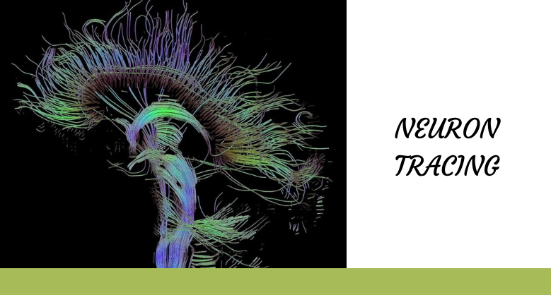
NEURON TRACING
Neuron Tracing or sometimes called Neuron Reconstruction.
The term itself is defined as the process of digitally reconstructing a whole neuron or some specific portions like the axon and dendrites of a neuron. Neuron reconstruction creates a digital and geographic model of neurons which help researchers with morphological analysis such as structure, length, segment, the diameter of neurons and also reconstruction of neurons in brain organisation, cell culture etc. The morphology and connectivity of neurons synapse with other neurons provide the working of neurons in the whole body. Neuron digital reconstruction or tracing began in the early days of neuroscience which is visualised under a light microscope and sometimes using an electron microscope.
You may come across why a scientist is so interested in reconstructing the neurons because it helps researchers to learn, memorise and see the behaviour pattern of neurons. Also used in neurodevelopmental, neurodegenerative, neuropsychiatric and neurological disorders like Alzheimer’s disease, schizophrenia, Parkinson’s disease, traumatic brain injury, Huntington’s disease etc.

Methods of reconstructing neurons–
There are few methods to reconstruct the neurons, they are-
1. Manually neuron tracing– Manually obtaining 2D or 3D images from the microscope and then after the software is used to import the image data and trace the segments of the neuron. It can be used from basic reconstruction drawing tools to highly specialised tools. The major drawback of manual tracing is it requires high labour work. Some open-source software used in manual tracing is Vaa3D.
2. Automated reconstructions-It is a powerful and necessary tool for rapid progress in detecting neuronal activity. Digital encoding enables both structural and functional data.
3. Semi-automated reconstructions– In this method reconstructing neurons does not require any previous image data from the microscope. Although, it requires a computer-controlled microscope-based system with software for neuron reconstruction tools to get a live camera image. This tracing gives out the best solution that is more acceptable, reduces time cost, and has good reconstruction accuracy.
There are various neuron tracing tools available. Some open-source tools are Vaa3D and its different modules, and commercial tools are Neurolucida, Neurolucida360, aivia etc. which are used by neuroscientists to trace and analyse neurons. New tools like ultra-tracer are launched which can trace arbitrarily large image volumes. It has been seen that Neurolucida is cited as over 7 times more widely used in neuron tracing than other available neuron tracing programs and has a more versatile system to produce a neuronal reconstruction.
Let’s talk about Neurolucida
Neurolucida
It is the first computer-assisted neuron reconstruction system specially designed for tracing neurons and generating morphological data from a historical specimen. Most trusted by neuroscientists around the world for understanding neurodegenerative diseases, and neuropathy and also for producing accurate anatomical models.
Key features-
- Creates highly accurate 2D-3D reconstructions
- Perform hundreds of quantitative morphometric analyses for the structure including metrics about neurons and groups of neurons.
- Reconstruct neurons from a single section or serial sections-we can reconstruct serial sections directly from a microscope or image by Neurolucida.
- Get started quickly and easily use the Neurolucida interface.
- Dedicated microscope hardware is used for efficient neuroanatomical data and image processing features like background correction, even and getting quality images using brightfield, darkfield and multi-channel fluorescence.
- Work with the image from other sources-we can reconstruct and analyse the imported image on any computer even when it is not connected to the microscope.
- Powerful quantitative analyses-Thousands of analysed parameter results can be exported from other software like R, Excel, MATLAB, and Python.
Latest Articles

International Women’s Day – Prioritising Women’s Health for a Healthier...
BY Ms. Sohini Chatterjee, Application Specialist, DSS Imagetech 7th March 2026
In 2013, one public disclosure transformed the global conversation around women’s health. When Angelina Jolie revealed in a New York Times op-ed that she carried a BRCA1 mutation and had...
Read More
How BX51WI Microscopes Support High-Resolution Neural Imaging
BY DSS Imagetech Pvt Ltd 24th February 2026
Picture this scenario. It’s 6:00 PM on a Friday. You have spent the last six hours harvesting tissue. You perfused the mouse perfectly, the liver cleared instantly, and the brain...
Read More
The Role of Research & Development in Driving Scientific Innovation
BY DSS Imagetech Pvt Ltd 24th February 2026
Human progress is not an accident. The leap from a simple medicinal herb to a targeted biologic drug, from a magnifying glass to a digital microscope, or from a basic...
Read More