DSS: Redefining Biotechnology & Life Science in India


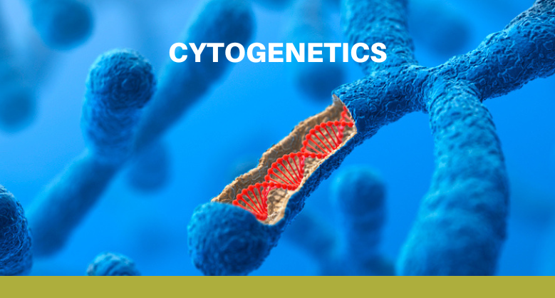
Cytogenetics
Cytogenetics is the branch of genetics that deals with the chromosome. Thus, this room is all about the chromosomes: chromosome structure and composition, the methods that scientists use to analyze chromosomes, chromosome abnormalities associated with disease, chromosomes play an important role in sex determination
Cytogenetics has been an important part of biology since 1842 when Swiss botanist Karl Nägeli first discovered the chromosomes in pollen. Since Nägeli’s discovery, methods for chromosome examination have become much more effective, further clarifying their roles in cell biology and human and animal health in ways undreamed of when chromosomes were first discovered. In 1870, German anatomist W. Flemming introduced aniline staining to visualize chromosomes during cell division for the first time.
In the decades since, this has been defined as the study of chromosomes, including their mechanics, behavior, and role in inheritance. Cytogenetics emerged in the early twentieth century when scientists observed that the chromosomes are the carriers of genes. As in science, researchers built on the observations of their fellow investigators to synthesize the chromosomal theory of heredity. In the early years of cytogenetics, scientists had a difficult time distinguishing the chromosomes, but over the years, they continued to refine the conditions for preserving and staining chromosomes to the reproducible standard that is now well noted in clinical cytogenetics. In today’s procedures, metaphase chromosomes are treated with stains that generate specific banding patterns, and chromosome pairs are then arranged into a precise format known as a Karyotype. Karyotypes are remarkably uniform i.e. species to species.
KARYOTYPE:
Karyotyping is the process of arranging or pairing all the chromosomes of an organism, thus providing a snapshot of an individual’s chromosomes. Karyotypes are prepared using standardized procedures that reveal characteristic structural features for each chromosome. A normal human contains 22 pairs of autosomes and 1 pair of sex chromosomes. Changes in chromosome number are easily detected in karyotypes. Cytogeneticists can then use coordinates on these chromosome maps, or ideograms, to identify the positions of structural abnormalities i.e. deletions, duplications, and translocations or Inversion. A most common aneuploidy is Down syndrome which has trisomy 21, which is frequently detected during prenatal screening. In fact, as medical genetics is well integrated with clinical medicine, karyotypes are becoming a source of diagnostic information for specific birth disorders, genetic disorders, and even cancers.
FISH:
Over the past few decades, Fluorescence in situ hybridization (FISH) has transformed cytogenetics into a molecular science and provided cytogeneticists with a powerful new tool to detect many chromosomal abnormalities. In FISH procedures, fluorescent-labelled DNA or RNA probes are hybridized with their complementary targeted DNA sequences on the chromosomes. FISH experiments generate coloured results because multiple probes are labelled with a spectrally distinct fluorescent dye. The target DNA sequences either have a single gene or a collection of genes along the length of a chromosome. FISH procedures are routinely emphasized in clinical cytogenetics. Spectral karyotyping(SKY) gives an overview of rearrangements and changes in chromosomes based on spectral microscopy. For SKY of human metaphase chromosomes, specific painting probes are used in just one FISH experiment. Using Locus specific, Chromosome enumeration probes cytogeneticists can also identify the genes or chromosomes that are affected by chromosomal mutations.
MICROARRAYS:
The current cutting edge of cytogenetics is microarrays. It uses microscope slides with thousands of tiny spots on which DNA or RNA probes are attached. The probes can be fluorescent-labeled or otherwise similar to the probes used in other techniques. By concentrating a probe, a single sample can be observed for thousands of different targets, and this technique can be adapted for many different kinds of probes, including single nucleotide polymorphisms (SNPs), copy number variations (CNVs), and more. The use of discrete wells in microarray data can be easily monitored, making for particularly rapid and thorough data collection.
Microarrays may replace all of their formers, but the future of cytogenetics isn’t in a single technique. Many more methods developed in the late 20th century are still relevant in the 21st. Techniques developed for genomics, such as Next-generation sequencing (NGS), also provide the same sorts of questions that cytogenetics does. Future innovations in cytogenetics will combine with the works that were all done in the past techniques to find upcoming, more efficient ways for cytogenetic data storage, as well as new ways for answering questions that are in the modern pipelines.
The collection of articles in this topic room is intended to provide an introduction to chromosome biology that has led to a basic understanding. Cytogenetics is a waste and growing field of research, and many topics have not been discussed in detail.
Latest Articles
How BX51WI Microscopes Support High-Resolution Neural Imaging
BY DSS Imagetech Pvt Ltd February 24, 2026
Picture this scenario. It’s 6:00 PM on a Friday. You have spent the last six hours harvesting tissue. You perfused the mouse perfectly, the liver cleared instantly, and the brain...
Read More
The Role of Research & Development in Driving Scientific Innovation
BY DSS Imagetech Pvt Ltd February 24, 2026
Human progress is not an accident. The leap from a simple medicinal herb to a targeted biologic drug, from a magnifying glass to a digital microscope, or from a basic...
Read More
Beyond the Microscope: Detecting Genomic Changes with D024 KaryoProfiler
BY DSS Imagetech Pvt Ltd February 17, 2026
In the Quiet Hours of the Lab In the quiet hours of a cytogenetics laboratory, cells are busy at work while no one is watching. They divide, adapt—and sometimes, silently,...
Read More