DSS: Redefining Biotechnology & Life Science in India


Spectral karyotyping
Spectral karyotyping (SKY) is a novel method for chromosome examination that has been created dependent on the methodology of the fluorescence in situ hybridization. Owing to its technology for painting each of the 24 human chromosomes with various colors, spectral karyotyping allows it possible to diagnose a range of disease [1].
Chromosomal analysis plays an important role in human leukemia diagnosis, treatment and prognosis [2-6]. In assessing the risk for phenotypic abnormalities, particularly in prenatal circumstances, the identification of marker chromosomes is significant. For the provision of information to couples about the possible phenotypic and/or developmental impact of a de novo rearrangement, the ability to determine the origin of additional genetic materials is very significant. Similarly, the detection of derivative chromosomal content will shed light on the mechanism of infertility when evaluating infertility [7-9]. In constitutional research, spectral karyotyping is an invaluable diagnostic tool for identifying marker chromosomes and chromosome exchanges that are not completely described by traditional cytogenetic methods and in identifying new subgroups of leukemia.
In this technology,chromosomes are painted with 24 chromosome-specific painting probes. This is called Combinatorial chromosome painting techniques. Using three fluorochromes and two haptens, the probes are labelled by degenerated oligonucleotide-primed PCR. Each probe is labeled differentially with one, two, three or four fluorescent dyes resulting in each chromosome having a special spectral signature. With similar principle of labelling the chromosomes,there emerged two techniques -Multiplex fluorescence in situ hybridization (mFISH) and Spectral Karyotyping (SKY), both follows similar basic fluorescence in situ hybridization (FISH) procedures, however the imaging methods are different [10].

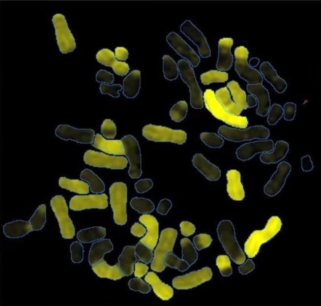
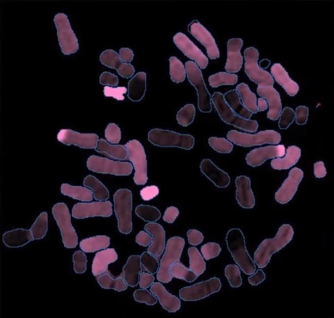
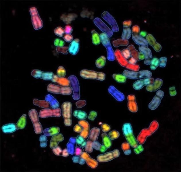
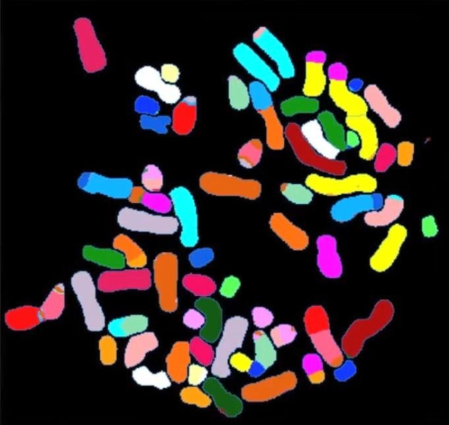
The differentiation of the chromosomes based on their spectral properties is defined by Spectral karyotyping (Fig1), while mFISH identifies the differentiation of the chromosomes based on the presence or absence of the fluorochrome when visualized with specific filters(Fig 2).

In SKY technology. using a traditional fluorescence light microscope fitted with a custom-designed triple-bandpass filter and the SpectraCube, a spectral image is acquired after in situ hybridization and immunodetection, which is able to retrieve spectral information for each pixel in a digital CCD image. The 24-colour display and chromosome classification are based on the chromosomes’ unique emission spectra. A detailed description of chromosomal aberrations is provided along with chromosome banding data from an inverted DAPI or a G-banded metaphase [11-12].

The center of the platform for SKY is a Sagnac Interferometer, installed on a rotatable disk to which a CCD camera is attached. The ray of light entering the triangle interferometer is divided into two beams. (a transmitted beam and a reflected beam), traveling in opposite directions, but in space on the same path. They’re hitting the CCD with Optical Path Difference (OPD) and shape a pattern of interference that is influenced in the image, by the spectral content at each point. It captures a sequence of images, at each different OPD, to form an interferogram at each pixel. A Fourier-analysis approach is applied to extract the sample’s hyperspectral image.
In MFISH, using specialized single band-pass filter sets and dedicated M-FISH software, microscopic visualization and digital acquisition of each fluorophore is obtained. These acquired images are then superimposed, allowing the classification of individual chromosomes in their group [13].
The main deficiency with mFISH is Cross talk between the filters. The emission spectrum of only two adjacent dyes is shown in the following example(Fig 5). Comparable filter-based measurements may provide fair results as long as the amplitude of those signals is comparable. However to compensate for its weak signal, if the green dye is faint, one will need to increase its exposure time. The cross talk factor will then be even more dominant than the green signal from other dyes (which spread all over due to 10-20 percent non-specific staining). This will cause large misclassifications.

GenASIs HyperSpectral platform is based on a cutting-edge, dual-mode optical device, which allows both interferometer-based image capture for hyperspectral imaging and direct view mode for high-resolution CCD image capture. The HyperSpectral data reveals the spectrum of every pixel in the image, and provides advanced analysis tools to extract quantitative and hidden information from within a sample. In Direct View mode, the system records image details under extremely low intensities and provides a finely detailed high resolution and high-definition image. SKY is hyper precise, accurate and hypersensitive.

References:
- Imataka, G. and Arisaka, O., 2012. Chromosome analysis using spectral karyotyping (SKY). Cell biochemistry and biophysics, 62(1), pp.13-17.
- Guo, B., Han, X., Wu, Z., Da, W. and Zhu, H., 2014. Spectral karyotyping: an unique technique for the detection of complex genomic rearrangements in leukemia. Translational pediatrics, 3(2), p.135.
- Mohr, B., Bornhäuser, M., Thiede, C., Schäkel, U., Schaich, M., Illmer, T., Pascheberg, U. and Ehninger, G., 2000. Comparison of spectral karyotyping and conventional cytogenetics in 39 patients with acute myeloid leukemia and myelodysplastic syndrome. Leukemia, 14(6), pp.1031-1038.
- Lu, X.Y., Harris, C.P., Cooley, L., Margolin, J., Steuber, P.C., Sheldon, M., Rao, P.H. and Lau, C.C., 2002. The utility of spectral karyotyping in the cytogenetic analysis of newly diagnosed pediatric acute lymphoblastic leukemia. Leukemia, 16(11), pp.2222-2227.
- Mrózek, K., 2008, August. Cytogenetic, molecular genetic, and clinical characteristics of acute myeloid leukemia with a complex karyotype. In Seminars in oncology (Vol. 35, No. 4, pp. 365-377). WB Saunders.
- Fadl‐elmula, I., Kytölä, S., Pan, Y., Lui, W.O., Derienzo, G., Forsberg, L., Mandahl, N., Gorunova, L., Bergerheim, U.S., Heim, S. and Larsson, C., 2001. Characterization of chromosomal abnormalities in uroepithelial carcinomas by G‐banding, spectral karyotyping and FISH analysis. International journal of cancer, 92(6), pp.824-831.
- Anguiano, A., Wang, B.T., Wang, S.R., Boyar, F.Z., Mahon, L.W., El Naggar, M.M., Kohn, P.H., Haddadin, M.H., Sulcova, V., Sbeiti, A.H. and Ayad, M.S., 2012. Spectral Karyotyping for identification of constitutional chromosomal abnormalities at a national reference laboratory. Molecular Cytogenetics, 5(1), p.3.
- Chen, C.P., Lin, C.C., Su, Y.N., Tsai, F.J., Chern, S.R., Lee, C.C., Chen, W.L., Chen, L.F., Wu, P.C. and Wang, W., 2010. Prenatal diagnosis and molecular cytogenetic characterization of a small supernumerary marker chromosome derived from chromosome 22. Taiwanese Journal of Obstetrics and Gynecology, 49(3), pp.381-384.
- Chen, C.P., Chen, M., Wu, C.H., Lin, C.J., Chern, S.R., Wu, P.S., Chen, Y.N., Chen, S.W., Chang, S.P., Chen, L.F. and Wang, W., 2017. Prenatal diagnosis and molecular cytogenetic characterization of mosaicism for a small supernumerary marker chromosome derived from chromosome 21q11. 2-q21. 1 and a literature review. Taiwanese Journal of Obstetrics and Gynecology, 56(4), pp.554-557.
- Loucas, B.D., 2019. Analysis of Radiation-Induced Chromosome Exchanges Using Combinatorial Chromosome Painting. In Radiation Cytogenetics (pp. 123-135). Humana, New York, NY.
- Macville, M., Veldman, T., Padilla-Nash, H., Wangsa, D., O’Brien, P., Schröck, E. and Ried, T., 1997. Spectral karyotyping, a 24-colour FISH technique for the identification of chromosomal rearrangements. Histochemistry and cell biology, 108(4-5), pp.299-305.
- Padilla-Nash, H.M., Barenboim-Stapleton, L., Difilippantonio, M.J. and Ried, T., 2006. Spectral karyotyping analysis of human and mouse chromosomes. Nature protocols, 1(6), p.3129.
- Anderson, R., 2010. Multiplex fluorescence in situ hybridization (M-FISH). In Fluorescence in situ Hybridization (FISH) (pp. 83-97). Humana Press, Totowa, NJ.
Latest Articles

LUNG CANCER CARE: THE POWER OF PRECISION DIAGNOSTICS & GENOMICS
BY Mr. Jaywant Chauhan, Application & Product Specialist, DSS Imagetech 19th May 2026
Lung cancer today is among the most common & serious types of cancer in the world. It happens when abnormal cells in the lungs begin to grow in a way...
Read More
How Digital Karyotyping Software Supports Prenatal Genetic Testing
BY DSS Imagetech Pvt Ltd 30th April 2026
It is a Tuesday morning. A couple is sitting in an OB-GYN office, holding hands so tightly their knuckles are white. They aren’t looking at their phones. They are staring...
Read More
National DNA Day 2026: Why DNA Matters for Modern Healthcare
BY Ms. Geddam Malini, Application & Product Specialist, DSS Imagetech 24th April 2026
Every cell in our body carries DNA, the code of life. April 25 marks DNA Day, it highlights advances in genomic research and encourages education in genetics. While DNA was...
Read More