DSS: Redefining Biotechnology & Life Science in India


Home > Microscopy & Imaging
Microscopy & Imaging
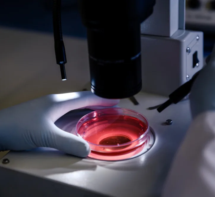
The Microscopy & Imaging division has been in existence since more than 40 years and is one of the foundation pillars of DSS. This long-standing experience in the business of Microscopy & Imaging has given the Microscopy & Imaging division a unique position as the most informed supplier of Microscopes and Digital Imaging solutions in India. As a complete solutions provider for microscope users across multiple user segments, DSS has developed unique skills and capabilities to configure and integrate Microscopy & Imaging based system solutions for life sciences research and clinical applications like Pathology IVF and Cytogenetics. To compliment the Microscopy & Imaging business DSS has successfully added to its portfolio a complete range of In-vivo imaging platforms.
The Microscopy & Imaging division ensures “Total Customer Satisfaction” by providing pan India support to our customers through our dedicated Application and Service teams.
The qualified and expert application team of Microscopy & Imaging is:
Dr. Anirban Bose has joined DSS Imagetech Pvt. Ltd. as a Product and Application Specialist for high-end imaging systems after completing his Ph.D. in Biochemistry from the University of Calcutta. His doctoral research concerns various biophysical techniques based on optical microscopy and spectroscopy and is conceded by a distinguished number of patents and publications in peer-reviewed journals. He also has expertise in developing autonomous optoelectronic sensing devices and their applications.
Ms. Nikita Kawli has done MSc (Applied Genetics), PGD MLT, PGD in Bio Nanotechnology She is working with DSS Imagetech as Product Application Manager covering a variety of Cytogenetics & Pathology Imaging products. Nikita has been involved in the entire cycle of Presales to post-sales activities. Professional competency in performing various assays on different samples, exposure to conducting various complicated tests to identify Infectious Diseases and Inheritable conditions, Calibration and Maintenance of various instruments, and Cytogenetics and Molecular Biological Works.
Dr. Shomnath Bhowmick is an expert in optics, material science, and advanced microscopy, with a strong focus on biological imaging and life sciences applications. Specializing in Confocal, STED, AFM, SEM, and TEM imaging, he has extensive experience in high-resolution characterization of biological and material systems. Holding a Ph.D. in Information Technology & Electrical Engineering (MEMS) from the University of Federico II, Naples, his research spans microfabrication, AFM-IR technique development, and bio-photonics. He has worked on MEMS resonators and thin-film characterization at SilTerra Malaysia and explored AFM, droplet microfluidics, and advanced imaging for biological applications at Shilps Sciences. Currently, as an Application Specialist & Sales Manager at DSS Imagetech, he provides technical expertise in AFM, Microfluidics, Confocal, Multiphoton, and STED microscopy, enabling cutting-edge research in cell biology, neuroscience, and bio-imaging. He holds multiple patents and has published extensively in leading scientific journals, contributing to advancements in biophysical imaging, nanoscale characterization, and photonics. His expertise extends to cleanroom processing, microelectronics, and computational modeling using COMSOL, MATLAB, and Python, bridging the gap between engineering, life sciences, and next-generation imaging technologies.
Dr. Sneha Paul completed her Ph.D. in Chemistry from University of Hyderabad, India. She is working with DSS Imagetech Pvt. Ltd. as an Application Specialist for high-end systems in the Micro Imaging Division. Prior to this, Sneha worked as a Postdoctoral Researcher at McGill University, Canada and University of Chicago, USA and as a Research Associate at CNRS-ISMO, France. She has expertise in advanced microscopic and spectroscopic techniques like confocal microscopy, super-resolution microscopy and transient absorption spectroscopy. She also has experience in developing image analysis algorithms.
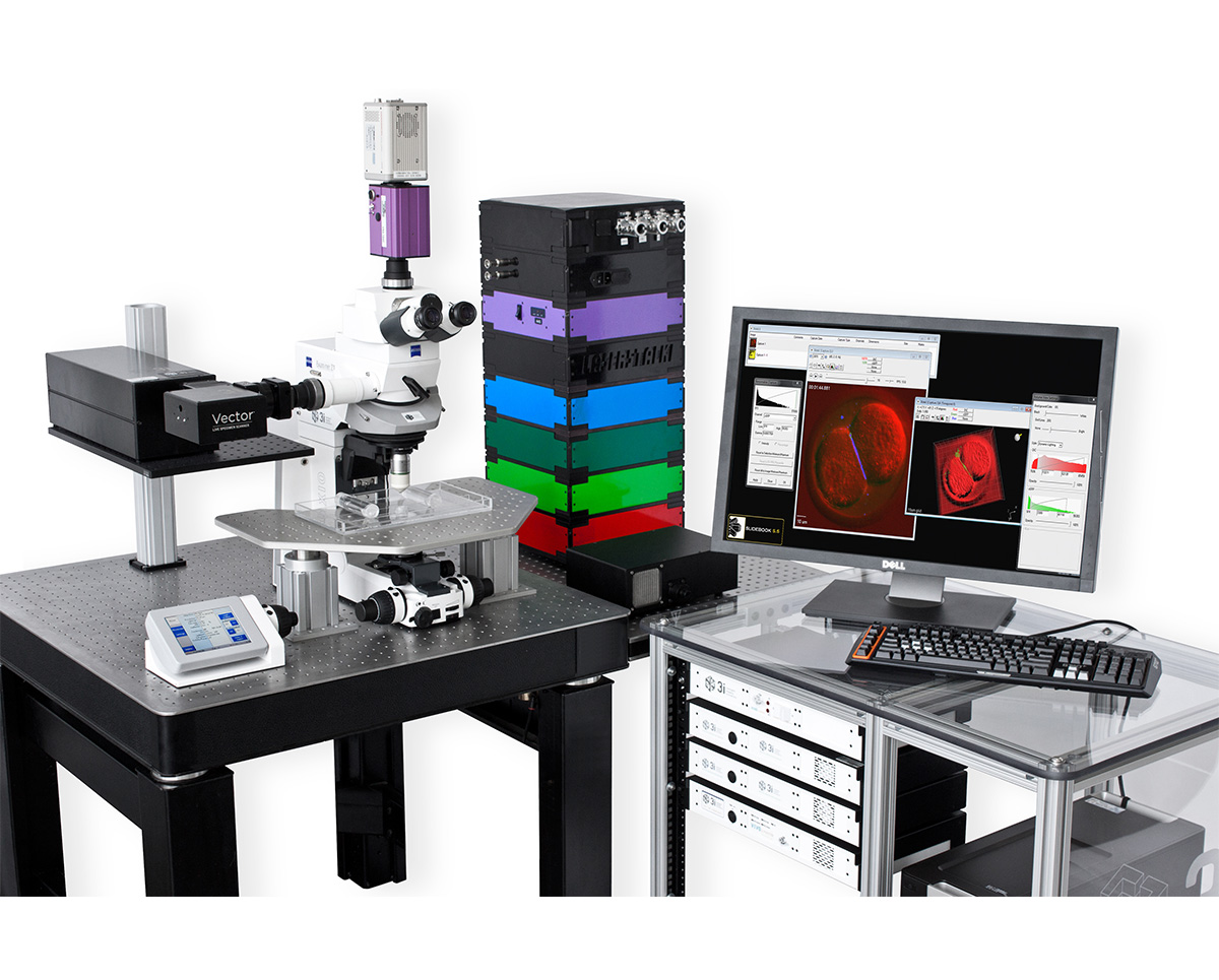
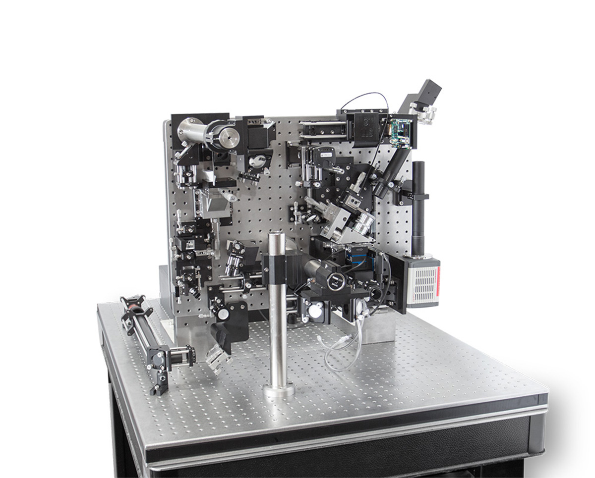
3i VIVO
VIVO incorporates advanced optics, cameras, computers and proprietary electronics to achieve unparalleled speed, precision and flexibility in live tissue image acquisition.
Applications & Specialities, Instruments, Microscopes, Microscopy, Products & Services, 3i, Microscopy & Imaging
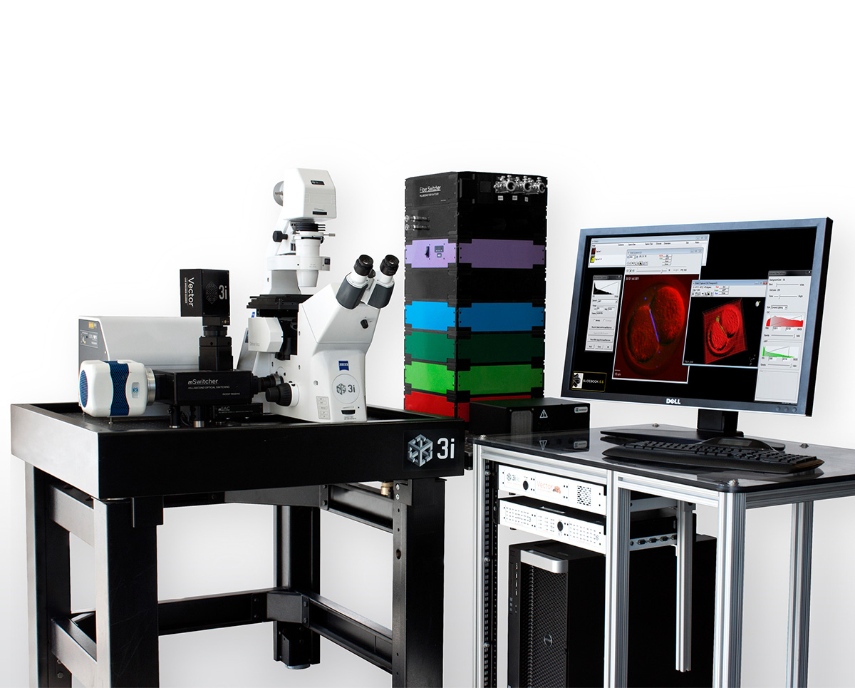
3i Marianas
Marianas incorporates advanced optics, cameras, computers and proprietary electronics to achieve unparalleled speed, precision and flexibility in live cell image acquisition.
Applications & Specialities, Instruments, Light Sheet, Microscopes, Microscopy, Products & Services, 3i, Microscopy & Imaging
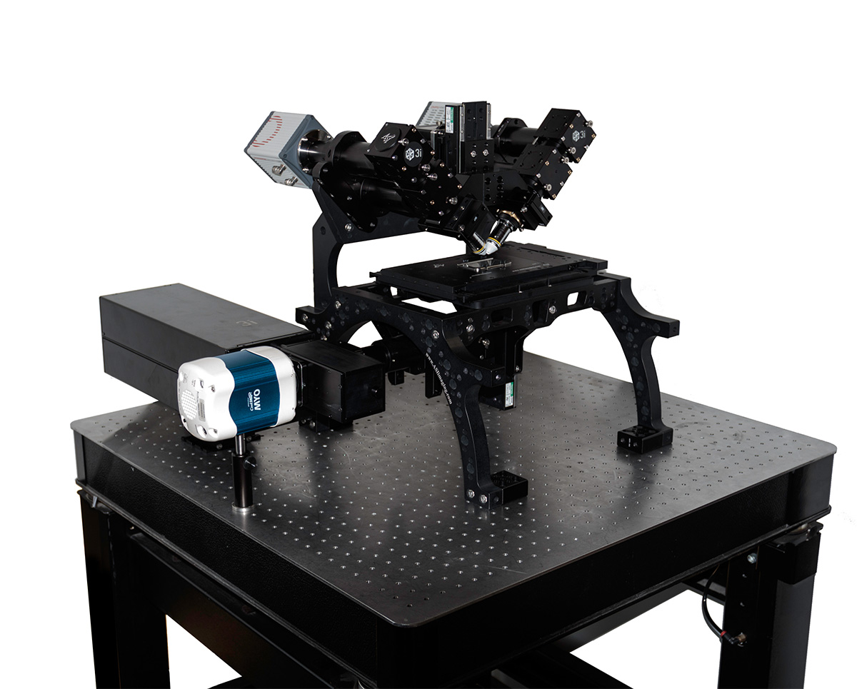
3i diSPIM
Dual-view Inverted Selective Plane Illumination (diSPIM) is a light sheet method offering low phototoxicity in 3D imaging making it possible to image living small organisms over extended periods of time.
Applications & Specialities, Instruments, Light Sheet, Microscopes, Microscopy, Products & Services, 3i, Microscopy & Imaging
3i Lattice LightSheet
Lattice LightSheet uses ultra-thin sheets of light to image 3D cellular dynamics for hundreds of volumes at dozens of frames per second at diffraction-limited resolution and super-resolution.
Applications & Specialities, Instruments, Light Sheet, Microscopes, Microscopy, Products & Services, 3i, Microscopy & Imaging
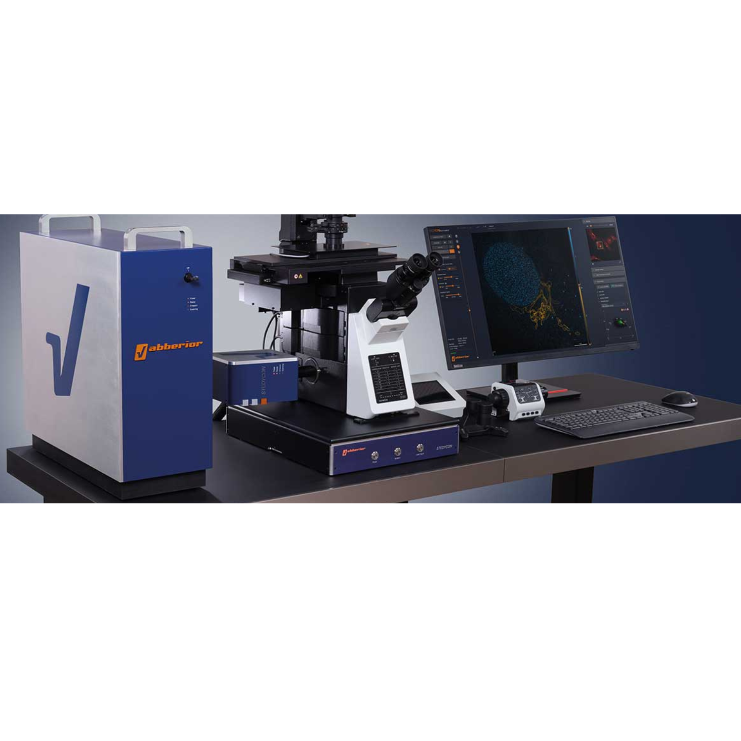
STEDYCON 2
The STEDYCON transforms your existing widefield microscope into an advanced confocal and STED nanoscope, achieving a remarkable resolution down to 30 nanometers. With just a free camera port and a quality objective lens, you can enhance your imaging capabilities significantly.
Applications & Specialities, Instruments, Microscopes, Products & Services, Super Resolution, Widefield, ABBERIOR INSTRUMENTS, Microscopy & Imaging

INFINITY
The INFINITY platform stands as the ultimate customisable solution for all microscopy applications. INFINITY is designed to meet every research challenge from STED and confocal microscopy to intravital imaging, material science, and optical trapping. Simply share your requirements, and we'll deliver a tailored, continuously upgradeable system precisely configured to support your research objectives.
Applications & Specialities, Instruments, Microscopes, Products & Services, Scientific Imaging, Widefield, ABBERIOR INSTRUMENTS, Microscopy & Imaging

FACILITY
FACILITY offers a revolutionary combination of advanced microscopy technology and user-friendly design, setting new standards for high-performance imaging. This system integrates state-of-the-art features ideal for both confocal and super-resolution imaging, all with intuitive, effortless operation.
Applications & Specialities, Instruments, Microscopes, Products & Services, Scientific Imaging, Super Resolution, ABBERIOR INSTRUMENTS, Microscopy & Imaging
Abberior STAR
Our abberior STAR dyes are designed to set a new benchmark in fluorescence light microscopy, offering unparalleled performance. These dyes boast extraordinary photostability and remarkable brightness, ensuring crisp, high-resolution imaging every time. Whether you are conducting STED or confocal imaging, our STAR dyes are the ultimate choice for achieving exceptional clarity and long-lasting fluorescence, making them a vital component of your research and imaging needs. There's no better way to elevate your microscopy results.
Applications & Specialities, Dyes, Kits Reagents & Consumables, Products & Services, Scientific Imaging, ABBERIOR INSTRUMENTS, Microscopy & Imaging

Abberior LIVE
Experience superior live-cell imaging with abberior LIVE dyes, meticulously crafted for STED (Stimulated Emission Depletion) and confocal microscopy. These innovative dyes set new benchmarks in live-cell labeling by delivering exceptional brightness and unmatched photostability. They simplify your research workflow, ensuring high-quality results every time.
Applications & Specialities, Dyes, Kits Reagents & Consumables, Products & Services, Scientific Imaging, ABBERIOR INSTRUMENTS, Microscopy & Imaging
Abberior FLUX
Our abberior FLUX dyes are specifically designed to provide unparalleled performance in MINFLUX imaging. Combining outstanding photostability with exceptional brightness, these dyes enable clear and accurate visualization while minimizing background noise. Their photoswitching kinetics are finely tuned for optimal results in single-molecule localization microscopy, ensuring precise and reliable data.
Applications & Specialities, Dyes, Kits Reagents & Consumables, Products & Services, Scientific Imaging, ABBERIOR INSTRUMENTS, Microscopy & Imaging
Abberior CAGE
abberior CAGE dyes represent a sophisticated family of fluorophores specifically engineered for advanced imaging techniques such as PALM, STORM, and GSD microscopy. These dyes exhibit a unique property: they remain non-fluorescent until activated by UV light. Upon exposure, they are transformed into intensely bright organic probes, delivering exceptional fluorescence with remarkable precision for localization. This uncaging mechanism makes them invaluable for applications requiring the highest levels of accuracy and brightness in single-molecule and super-resolution microscopy, significantly enhancing imaging outcomes and data reliability.
Applications & Specialities, Dyes, Kits Reagents & Consumables, Products & Services, Scientific Imaging, ABBERIOR INSTRUMENTS, Microscopy & Imaging
Abberior Supplies
Ready-to-Image Alignment Sets - abberior ALIGNMENT SETS simplify the calibration and testing of fluorescence microscopes. Each set includes multiple microscope slides pre-prepared with nanoparticles or cell specimens, allowing for immediate imaging and precise adjustments.
Applications & Specialities, Dyes, Kits Reagents & Consumables, Products & Services, Scientific Imaging, ABBERIOR INSTRUMENTS, Microscopy & Imaging
Testimonials & Reviews
Dr. (Prof.), Nitesh Mohan
Professor & Head, Department of Pathology, RMCH Bareilly
DSS's expertise, dedication, and professionalism were outstanding in making the Karyotyping & FISH workshop a great success. Their knowledge and valuable insights empowered all the participants with practical skills, receiving highly positive feedback from both students as well as faculty members.

Dr. Chhaya Chande, Professor & HOD, Microbiology
GGMCJJ Hospitals, Mumbai
“Ms. Megha Dhumal (Assistant Manager- Application) has done a satisfactory demonstration of the running of the Abbott Sample preparation machine model m2000sp and the Abbott RT-PCR machine model m2000rt. We appreciate the effort made by the DSS team under these difficult conditions to help our lab to carry out the imperative Covid-19 tests.”
Dr Sunil K Arora, Professor, Deptt of Immunopathology
PGIMER, Chandigarh
“We are using Confocal Microscope and one Fluorescence Microscope. Both are working fine. The after sales services by DSS have been excellent for functioning & upkeep of the microscopes. The applications support by experts from DSS is very useful. Keep it up!”
Dr Pramod Kumar Bajaj
MD, Spermprocessor Pvt Ltd
“Really excited to see the DSS Pathology solutions exhibition booth at APCON 2019 along with Magnus. We think all the upcoming technology had been displayed along with your efforts to make it Indigenous (Made in India) is highly appreciated. Wish you all the best. Keep it up!”
Dr. Sreejesh S, Associate Professor, Dept of Hematology
PGIMER, Chandigarh
“My experience with DSS so far has been very good till now. We are getting good support in both purchase as well as in troubleshooting. Very good experience with Mr Arun, Mr Manoj, Mr Mahesh and all others from the DSS team.”
Dr Sudha S Murthy, Department of Pathology and Laboratory Medicine
BIACH & RI, Hyderabad
“I am happy with DSS and associated with 19 years and use Dako antibody. Happy with Supply but need improvement.”
Dr S Radhika MD, PhD
Professor, Deptt. Of Cytology & Gynaec Pathology, PGIMER, Chandigarh
“PGI Cytology Dept. has had a long association with DSS- Olympus Microscopy Division. They have provided excellent services- after sales service. The product is also of very good quality. We have had no problems with their products and services are of very good quality.”
Dr Nuzhat Husain
RMLIMS, Lucknow
“Have been using Dako Reagents and Dako antibodies for a while. Services and products have been good and timely.”
Dr Minu Singh
Assistant Professor, PGIMER, Chandigarh
“MRC Holland MLPA products provided by DSS are of good quality, have never faced any quality issues with their product or shipping condition. They provide prompt response upon any query.”
Mr. Krishnani Professor, SGPGI, Lucknow
“My experience with DSS so far has been excellent for the last 30 years- sales and service experience. Microscope products are very useful and sturdy with high precision.”