DSS: Redefining Biotechnology & Life Science in India


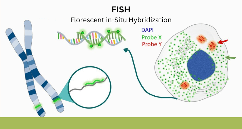
FISH
Don’t be confused with the fish you know. Here, FISH stands for florescent in-situ hybridisation. It is a molecular cytogenic technique which uses florescent probe that binds to only those segments which have high degree of sequence complementarity. In early 1980s it was developed by biomedical researchers to determine the presence or absence of DNA or RNA sequences on chromosomes, as well as to localize these DNA sequences on metaphase chromosome. It is a genetic technique used to diagnose congenital diseases and also been used to detect cancer and to diagnose other infectious diseases.
PRINCIPLE OF FISH
The basic elements of FISH are a DNA probe and a target sequence. Before hybridization, the DNA probe labelled either directly through incorporation by fluorophore or indirectly with non-fluorescent hapten (Haptens are small-molecular-weight compounds that evoke an immune response only when they are attached to carrier proteins). The labelled probe and the target DNA are denatured and the denatured probe and target DNA allows the annealing due complementary of DNA sequences. If the probe has been labelled indirectly, an extra step is required for visualization of the nonfluorescent hapten that uses fluorescent labelled antibody against hapten. Whereas FISH is faster with directly labelled probes. Finally, the signals are evaluated by fluorescence microscopy.

TYPES OF PROBE
PROBE is a nucleic acid which is labelled with marker for identification and will hybridisize with another nucleic acid on base complemetarity.
- Chromosomal painting or whole chromosome paintaing(wcp)- It refers to hybridisation of fluorecently labelled DNA probe to characterise choromosome rearrangements.Probe paint the entire length of chrmosome or any part of target chromosome with different color to get banding pattern and also to identify chromosomal aberrations. It is limitted to metaphase analysis as signal get diffuse in interphase.
- Gene-specific or locus-specific probes are derived from unique DNA sequences or loci within the chromosome which vary in size from 1-10kb in plasmid cloning vector to 100-300kb in P1 bacteriophage. These probes are very useful for detection of translocations, inversions, and deletions in both metaphase and interphase and also very useful in gene mapping.
- Telomeric or Centromeric repetitive sequence probes- These are generated from repetitive sequence of TTAGGG at the end of the chromosome and also found in the centromeres. Centromeric probes target the α- and β-satellite sequences. These centromere-specific probes are useful in detection of monosomy, trisomy, and other aneuploidies in tumours.
APPLICATION OF FISH
FISH has become an important tool for detecting chromosomal abbreviation and gene mapping.
- Parental diagnosis- FISH is used to detect congenital disease such as down’s syndrome (presence of an extra chromosome 21), Edward’s syndrome (presence of an extra copy of chromosome 18) etc.
- Detection of copy number variants (CNVs) -FISH have higher resolution than gimesa stain to detect genetic diseases. It takes less time to visualize structural features of chromosomes and help to detect chromosomal deletions, translocation etc.
- Cancer cytogenetics – FISH is rapid and highly sensitive in detecting cancer. FISH directly on tumor samples help to detect chromosomal aberration without the need of interphase chromosome. Recently FISH test is done on breast cancer which is removed during biopsy to detect extra copies of HER2 gene.
- Bacterial pathogen identification- FISH helps in diagnosis of infectious disease by taking sample from infectious patient. Pathogen cells will show colour if the pathogen is present.
- Fish also helps to deduce evolutionary disease.
- Interphase FISH is used in bone marrow transplantation.
Application of FISH in increased due to its accuracy, reliability, high resolution, and straightforward in comparison to other techniques. It also supports large-scale mapping in human genome project. Hence FISH should be preferred approach in gene expression leading to any disease.
Latest Articles

LUNG CANCER CARE: THE POWER OF PRECISION DIAGNOSTICS & GENOMICS
BY Mr. Jaywant Chauhan, Application & Product Specialist, DSS Imagetech 19th May 2026
Lung cancer today is among the most common & serious types of cancer in the world. It happens when abnormal cells in the lungs begin to grow in a way...
Read More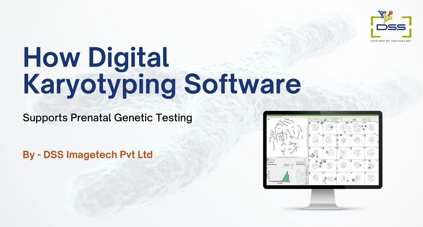
How Digital Karyotyping Software Supports Prenatal Genetic Testing
BY DSS Imagetech Pvt Ltd 30th April 2026
It is a Tuesday morning. A couple is sitting in an OB-GYN office, holding hands so tightly their knuckles are white. They aren’t looking at their phones. They are staring...
Read More
National DNA Day 2026: Why DNA Matters for Modern Healthcare
BY Ms. Geddam Malini, Application & Product Specialist, DSS Imagetech 24th April 2026
Every cell in our body carries DNA, the code of life. April 25 marks DNA Day, it highlights advances in genomic research and encourages education in genetics. While DNA was...
Read More