DSS: Redefining Biotechnology & Life Science in India
























Think Fungal: The Infection We Often Miss
Fungal Disease Awareness Week | 14–18 September 2026
2026 marks the 10th Annual Fungal Disease Awareness Week, highlighting a decade of efforts to raise awareness of fungal diseases and their impact.
A patient has persistent fever. Antibiotics have already been started, but the chest X-ray is inconclusive and routine bacterial cultures have not provided an answer.
A new specimen arrives in the laboratory microscope. The usual possibilities have been investigated, but there is still no clear explanation.
At this point, one question can change the direction of the investigation:
Could this be a fungal infection?
Fungi can be difficult to detect because they may be present at low abundance, embedded within tissue, slow-growing, or difficult to distinguish by morphology alone.
When the usual answers do not fit, think fungal.
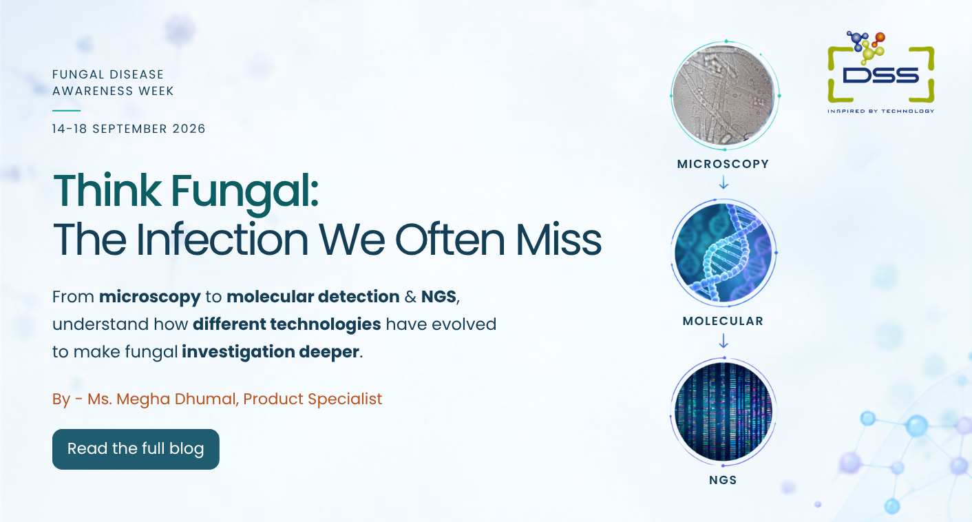
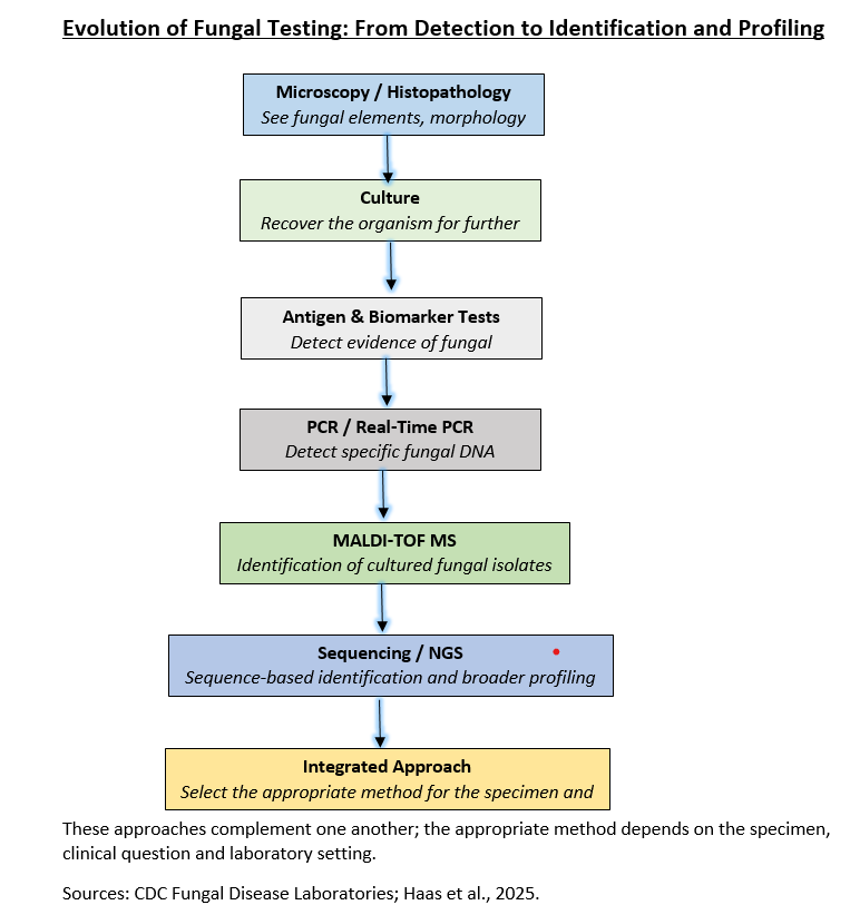
Depending on the clinical question, investigation may extend from microscopy and culture to molecular methods and sequencing – from seeing fungal structures to identifying and profiling the organism.
What Makes Fungi Different?
Fungi are eukaryotic organisms that occur in forms such as yeasts and moulds. Some occur as single-celled yeasts, while others grow as filamentous structures called hyphae, which can form a network known as mycelium. Some fungi can also change their form depending on environmental conditions.
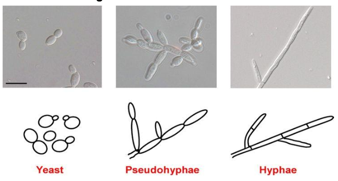
Image Credit: Childers et al. (2011) / ResearchGate
These differences matter in the laboratory. Fungi may grow slowly, occur at low abundance, or be difficult to distinguish based on morphology alone.
Fungi are not simply another type of bacteria. Their biology, growth patterns and interaction with the host are different – and so are the challenges involved in detecting them.
A World That Has Always Been Full of Fungi
Fungi are widespread in soil, water, plants, food, animals and the built environment. What has changed is the range of conditions in which humans and fungi interact.
Advances in transplantation, cancer treatment, intensive care and immunosuppressive therapies have increased the number of people vulnerable to opportunistic fungal infections. At the same time, fungal pathogens are emerging in new settings and developing resistance to available antifungal therapies.
In 2022, the World Health Organization published its first Fungal Priority Pathogens List, identifying 19 fungi of major public-health importance and highlighting the need for stronger surveillance, research and diagnostic capacity.
Fungal disease is no longer a niche concern.
The Fungal Threat Is Changing
Fungal infections can range from superficial or mucosal disease to deep-seated and invasive infections involving the lungs, bloodstream, brain or other organs. While many fungal exposures do not cause illness, invasive infections can become severe or life-threatening, particularly in people with weakened immune systems.
Candida auris has emerged as an important healthcare-associated pathogen, with some strains showing multidrug resistance. Aspergillus fumigatus, a common environmental mould, can cause severe disease in vulnerable patients, while azole-resistant strains are an increasing concern.
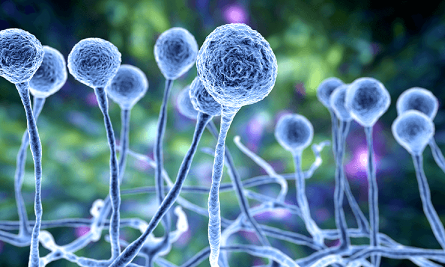
Source: https://www.news-medical.net/life-sciences/What-are-Fungi.aspx
In India, difficult-to-treat dermatophyte infections, including those associated with Trichophyton indotineae, have added another dimension to the fungal disease landscape.
These examples highlight a common challenge: laboratories need methods that can support timely identification while keeping pace with a changing fungal threat.
India: Where the Fungal Story Becomes Impossible to Ignore
The COVID-19 pandemic brought the impact of fungal disease in India into sharp focus.
During the major COVID-19 waves, particularly in 2021, India experienced a large increase in COVID-associated mucormycosis, especially among patients with diabetes and those exposed to corticosteroids. The outbreak highlighted how quickly fungal infection can become a major public-health concern when risk factors, clinical awareness and laboratory preparedness intersect.
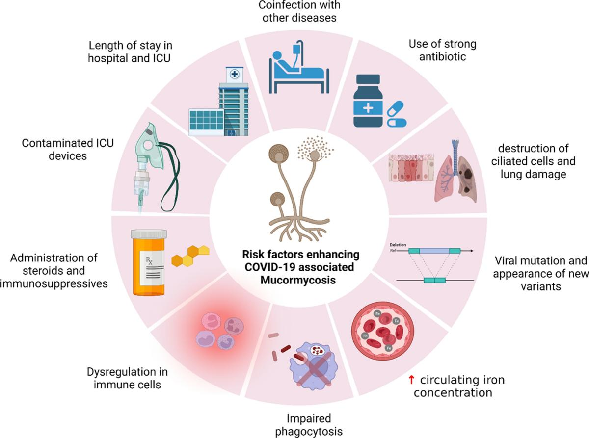
Source: https://pmc.ncbi.nlm.nih.gov/articles/PMC10516834/
But mucormycosis is only one part of the picture. Aspergillus, Candida, Cryptococcus, Histoplasma, Sporothrix and dermatophytes contribute to a much broader spectrum of fungal disease.
A 2022 modelling study estimated that more than 57 million people in India could be affected by serious fungal diseases each year. This is a modelled estimate rather than a complete national surveillance figure, but it illustrates the potential scale of the problem.
If fungal disease is not considered, it may not be tested for.
The Laboratory Story: First, See It
Once fungal disease enters the differential diagnosis, the laboratory becomes central to the investigation.
No single method answers every fungal question. The appropriate approach depends on the specimen, clinical context, suspected organism and information required.
Microscopy: The First Clue
Microscopy can provide rapid evidence of fungal structures, while histopathology can reveal fungal elements within tissue and help demonstrate tissue invasion. Stains such as Periodic acid–Schiff (PAS) and Grocott’s methenamine silver (GMS) can enhance the visibility of fungal structures.
However, low fungal burden, poor specimen quality, overlapping morphology and observer experience can affect interpretation.
Microscopy can provide the first clue – but the clue may need to be investigated further.
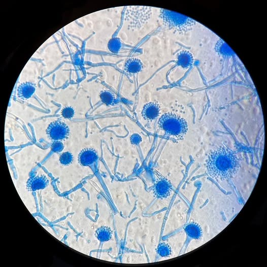
Aspergillus sp. isolated from soil and stained with lactophenol cotton blue. Image courtesy of Microbe Notes
Then, Grow It
Culture remains an important part of conventional mycology because it can recover the organism and, when a suitable isolate is obtained, support identification and antifungal susceptibility testing.
Some fungi grow slowly or are difficult to recover, and a negative culture does not necessarily exclude invasive fungal disease.
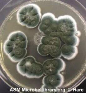
Colony morphology of Penicillium spp. grown on Sabouraud Dextrose Agar. Image courtesy of Hare / ASM MicrobeLibrary
Once an isolate is recovered, identification may also be supported by methods such as MALDI-TOF mass spectrometry, depending on the organism, database and validated laboratory workflow.
Some fungal pathogens also present specific challenges for conventional culture. For example, Pneumocystis jirovecii cannot be propagated in routine culture, making microscopic and molecular approaches particularly important for its detection.
Antigen and biomarker-based methods, including galactomannan and β-D-glucan in appropriate clinical settings, can provide additional evidence.
The next question becomes:
Can we look for the organism at the level of its genetic material?
The Molecular Approach
PCR provides another way to investigate suspected fungal infection by detecting specific fungal DNA directly from appropriate clinical specimens, without requiring prior culture growth. However, molecular testing is most useful when there is a defined diagnostic question and should be interpreted alongside other laboratory and clinical findings.
When the Target Is Known, Go Focused
When a particular fungal pathogen is suspected, targeted PCR can provide a focused investigation.
For example, the Certest VIASURE Aspergillus Differentiation Real-Time PCR assay is designed for the qualitative detection and differentiation of Aspergillus fumigatus, A. flavus and A. terreus DNA from respiratory specimens such as BAL, BAS and sputum in individuals suspected of aspergillosis.
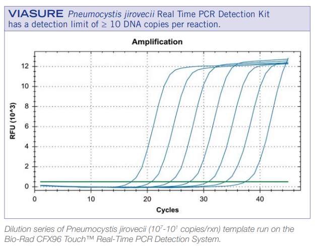
Similarly, the Certest VIASURE Pneumocystis jirovecii Real-Time PCR Detection Kit enables qualitative and quantitative detection of Pneumocystis jirovecii in respiratory samples.
For Candida auris, the Certest VIASURE Candida auris Real Time PCR Detection Reagents (RUO) provide a targeted molecular approach for the qualitative detection of C. auris DNA by targeting a conserved region of the ITS2 gene.
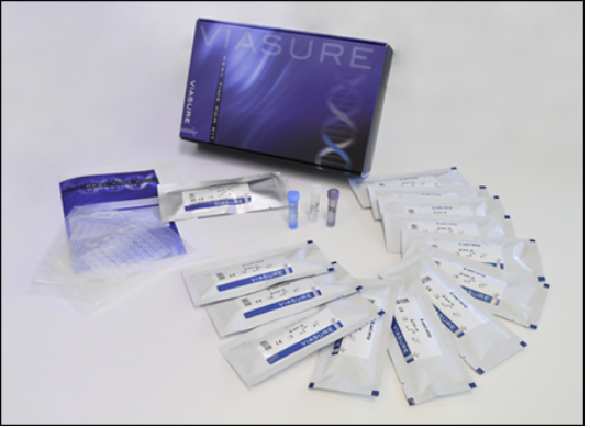
The VIASURE Real-Time PCR portfolio uses lyophilized, ready-to-use reagents designed for convenient handling, with room-temperature storage and transport and a shelf life of up to 24 months, depending on the product.
These examples illustrate how targeted molecular testing can complement conventional approaches when there is a specific organism or clinical question in mind. It does not replace microscopy, culture or other laboratory methods.
Importantly, detection of fungal DNA does not, by itself, establish active fungal disease. Results should be interpreted alongside the specimen type, clinical findings and other laboratory evidence. Depending on the organism and specimen, colonisation, contamination and true infection may need to be distinguished.
Microscopy shows structure. Culture supports recovery. PCR detects a specific genetic target.
But What If We Do Not Know What We Are Looking For?
Targeted PCR works best when there is a defined question.
But sometimes the laboratory faces a broader one:
What bacterial and fungal organisms may be present in this specimen?
This is where sequencing can provide a wider view.
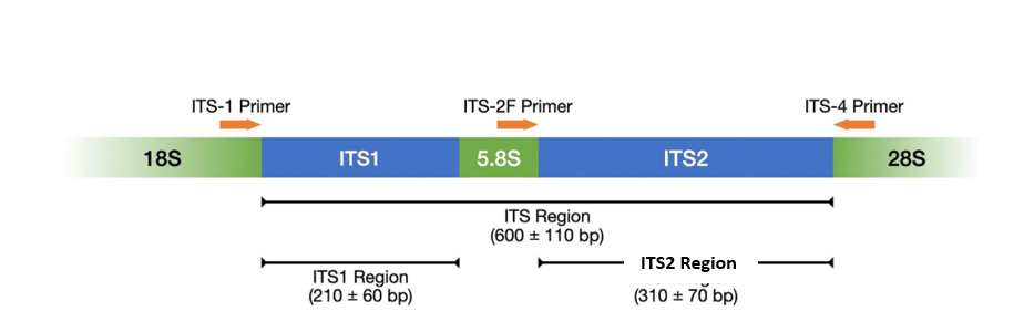
The Internal Transcribed Spacer (ITS) region is widely used for fungal identification and taxonomic profiling. ITS2, in particular, is commonly used for fungal taxonomic profiling, although the level of identification depends on the organism, specimen quality, assay design and reference database.
From One Target to a Broader View
The VIASURE Bacterial & Fungal NGS Solution (RUO) uses targeted amplification of bacterial 16S V1–V3 and fungal ITS2 regions to support simultaneous bacterial and fungal DNA profiling, followed by sequencing and bioinformatic analysis through V-Xplora. The solution uses ready-to-use, lyophilized reagents designed for room-temperature storage and transport, with a shelf life of up to 24 months.
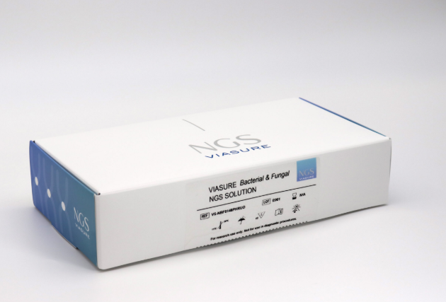
When the target is known, a focused approach may be appropriate. When the question is broader, targeted sequencing can help provide a wider microbial view. NGS is not required for every fungal investigation, but it can provide additional information when a broader microbial profile is needed.
Finding the Fungus Is Only Part of the Story
Identification is an important step, but it is not always the end of the investigation.
Antifungal approaches include polyenes, azoles and echinocandins, depending on the organism, site of infection and clinical context.
The challenge becomes greater when fungi develop antifungal resistance. Azole-resistant Aspergillus fumigatus, multidrug-resistant Candida auris and difficult-to-treat dermatophyte infections highlight the importance of timely identification and, where appropriate, susceptibility testing.
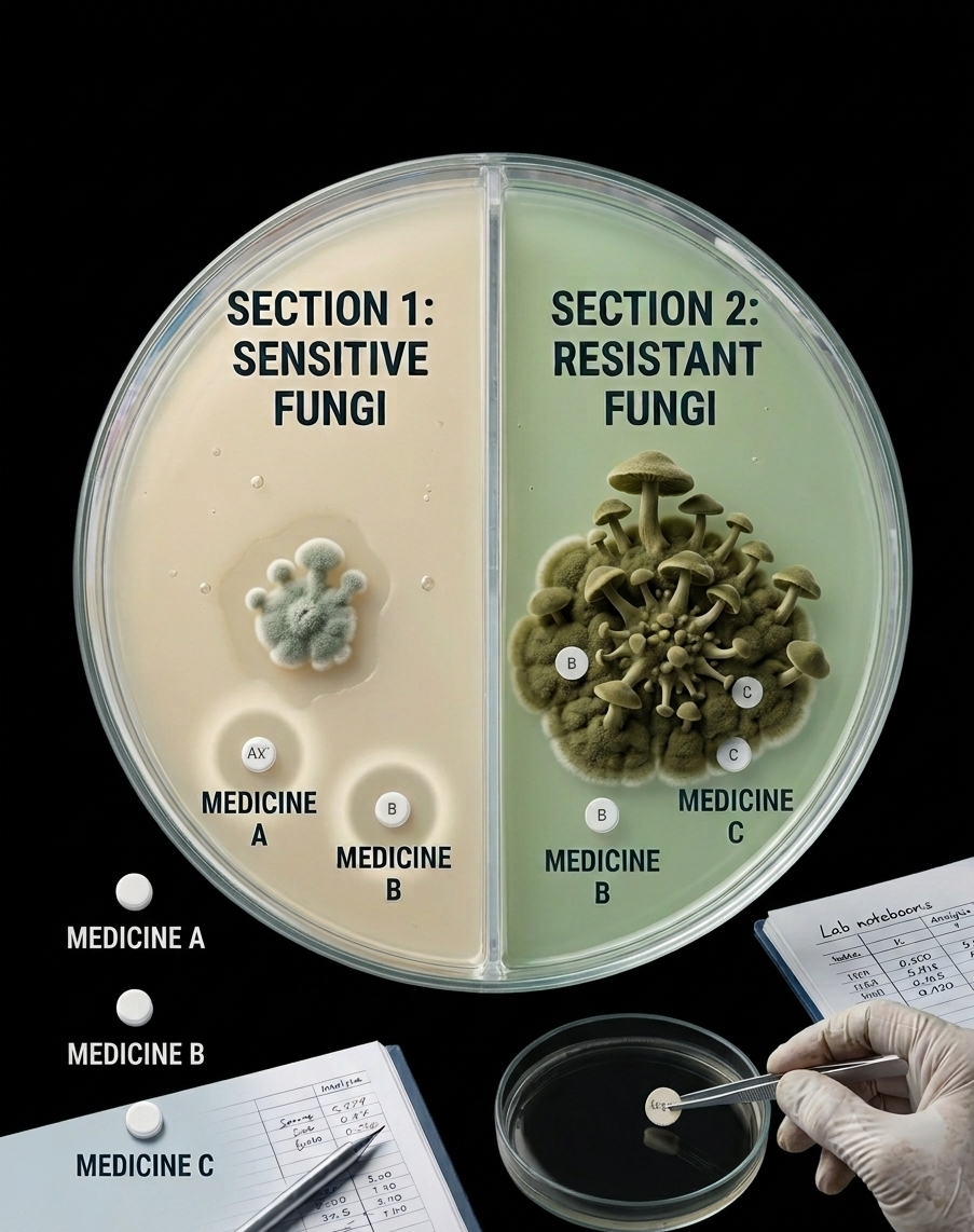
Culture-based susceptibility testing remains important. Molecular and genomic approaches can provide additional information about resistance-associated changes and, in appropriate settings, help investigate transmission and relatedness.
Knowing which fungus is present is often the first step towards understanding how the infection needs to be investigated further.
Fungi Do Not Stop at the Hospital Door
Fungi exist across interconnected environments — humans, animals, plants, soil, water, agriculture and the built environment.
Changes in climate, environmental conditions, healthcare practices and antifungal use can influence this landscape. Understanding fungal disease therefore increasingly benefits from a One Health perspective, where human, animal and environmental health are considered together.
The factors influencing fungal emergence and resistance can extend beyond a single patient, hospital or laboratory.
From Seeing to Understanding
The story of fungal investigation is not about replacing one technology with another.
It is about asking better questions at each stage.
A microscope may reveal the first clue. Culture may help recover the organism. Targeted PCR can provide a focused molecular answer. Sequencing can offer a broader view when the organism is uncertain or the microbial picture is complex.
The value lies in choosing the right tool for the right question.
Microscopy can provide an important first step in mycological investigation. DSS microscopy solutions, including Evident (formerly Olympus) & Magnus systems, provide platforms for examining fungal structures and other microscopic findings as part of the laboratory workflow. When microscopy raises suspicion, further investigation may include culture, antigen or biomarker testing, targeted molecular methods or sequencing, depending on the clinical question.
The Question We Should Leave With
Fungal disease can be difficult to see, difficult to grow and sometimes difficult to identify.
Yet the laboratory toolbox has evolved considerably – from microscopy and histopathology to culture, antigen and biomarker testing, PCR and sequencing.
Technology can provide powerful answers, but only when the right question is asked.
Awareness begins with recognising that a fungal infection is possible, especially when the usual answers do not fit.
Think Fungal.Uncover the Fungal Signature.
FAQ’s :-
1. Why can fungal infections be difficult to diagnose?
Fungi may be present at low levels, grow slowly, or be difficult to distinguish by morphology alone, making laboratory detection challenging.
2. When should a fungal infection be considered?
Fungal infection should be considered when symptoms persist, routine investigations do not provide a clear explanation, or clinical risk factors raise suspicion of fungal disease.
3. What role does Real-Time PCR play in fungal detection?
Targeted Real-Time PCR can detect specific fungal genetic targets directly from appropriate specimens and complement conventional methods such as microscopy and culture.
4. When can NGS be useful for fungal investigation?
NGS can provide a broader microbial view when the causative organism is uncertain or when both bacterial and fungal organisms need to be investigated. The VIASURE Bacterial & Fungal NGS Solution (RUO) targets bacterial 16S V1–V3 and fungal ITS2 regions for microbial DNA profiling.
5. Can molecular testing replace conventional fungal methods?
No. Microscopy, culture, antigen or biomarker testing, Real-Time PCR and sequencing provide different types of information. The appropriate approach depends on the specimen, suspected organism and clinical question.
References
Centers for Disease Control and Prevention (CDC). Fungal Disease Awareness Week 2026. CDC – Fungal Disease Awareness Week
World Health Organization (WHO). WHO Fungal Priority Pathogens List to Guide Research, Development and Public Health Action. 2022. WHO – Fungal Priority Pathogens List
Ray A, Aayilliath K, Banerjee S, Chakrabarti A, Denning DW. Burden of Serious Fungal Infections in India. Open Forum Infectious Diseases. 2022;9(12). PubMed – Burden of Serious Fungal Infections in India
Singh AK, Singh R, Joshi SR, Misra A. Mucormycosis in COVID-19: A systematic review of cases reported worldwide and in India. Diabetes & Metabolic Syndrome: Clinical Research & Reviews. 2021. PMC – Mucormycosis in COVID-19
Centers for Disease Control and Prevention (CDC). Clinical Overview of Candida auris. 2026. CDC – Clinical Overview of Candida auris
Uhrlaß S, Verma SB, Gräser Y, et al. Trichophyton indotineae—An Emerging Pathogen Causing Recalcitrant Dermatophytoses in India and Worldwide—A Multidimensional Perspective. Journal of Fungi. 2022;8(7):757. PubMed – Trichophyton indotineae

About the Author
Ms. Megha Dhumal is a Product Specialist – Integrated Diagnostics, with 8+ years of experience in molecular diagnostics. She specializes in hematologic malignancy testing using real-time PCR, capillary electrophoresis, and NGS, with expertise spanning across solid tumors, genetic disorders, and infectious diseases. In her current role, she supports laboratories in implementing and optimizing integrated molecular diagnostic workflows, enabling accurate and reliable molecular results.
Latest Articles
Think Fungal: The Infection We Often Miss
BY Megha Dhumal, Product Specialist 18th September 2026
Fungal Disease Awareness Week | 14–18 September 2026 2026 marks the 10th Annual Fungal Disease Awareness Week, highlighting a decade of efforts to raise awareness of fungal diseases and their...
Read More
Super-Resolution Microscopy: Upgrading to Abberior STEDYCON for Advanced Research
BY Dr. Anirban Bose (Ph.D.) Product & Application, Manager 5th September 2026
No matter how high-end your conventional fluorescence or confocal microscope is, there is a hard ceiling that eventually becomes incredibly frustrating. You could be trying to get a clear look...
Read More
How to Choose the Right Research Microscope
BY DSS Imagetech Pvt Ltd 14th August 2026
You’re standing there with a procurement form and a budget number. Someone circled in red, and down the hall a colleague is muttering that their old scope “still works fine,...
Read More