DSS: Redefining Biotechnology & Life Science in India


















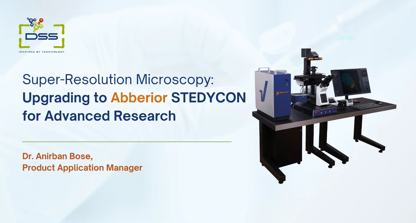
Super-Resolution Microscopy: Upgrading to Abberior STEDYCON for Advanced Research
No matter how high-end your conventional fluorescence or confocal microscope is, there is a hard ceiling that eventually becomes incredibly frustrating.
You could be trying to get a clear look at sub-organelle structures, synaptic protein clusters, a mitochondrial membrane, or a nuclear pore complex, but the details are constantly blurred together by the fundamental physics of light itself.
Once you hit that wall, no amount of increasing laser power, optimizing contrast, or refining detector sensitivity can recover the lost high-spatial-frequency information; the bottleneck is not engineering but wave optics itself.
What you’re running into is the classic diffraction limit. Discovered as far back as 1873, this principle essentially states that you cannot resolve two distinct points if they are closer than roughly half the wavelength of the light passing through the lens.
In reality, once the feature dimensions fall below the ~170–250 nm set by Abbe’s limit, the details are irretrievably blurred together by the point-spread function of the far-field optical system.
So now, studying cell biology, protein clustering, or membrane architecture, a roughly 200 nm limit is a significant roadblock. The actual biological interaction you want to see is happening at scales of 20, 50, or 80 nm; the smaller the features, the harder to reveal.
Your standard microscope may simply show a blurry blob of emitted light where the real story is taking place.
Super-resolution microscopy was developed to break through this barrier.
The Abberior STEDYCON represents one of the most practical, accessible, and technically refined approaches to achieving this in a format that is increasingly gaining traction in Indian research institutions.
A Quick Primer on Super-Resolution: What Are We Actually Talking About?
Before diving into the STEDYCON itself, it is worth taking a step back to look at the broader super-resolution landscape, particularly because these techniques are not all created equal.
STORM, PALM & DNA-PAINT
Take STORM, PALM & DNA-PAINT, for example, the basic mechanism of single-molecule localization methods works by randomly switching individual fluorescent molecules on and off and gradually reconstructing a super-resolved image from thousands of snapshots over time.
While they can deliver remarkable localization precision of below 20 nm, they are relatively slow, require specific fluorophores and sample preparation, and involve substantial computational post-processing with lots of optimization.
Therefore, for real-time live-cell applications, their use can be challenging because they are primarily suited to fixed and relatively static samples.
Structured Illumination Microscopy (SIM)
On the other hand, there is Structured Illumination Microscopy (SIM). By projecting patterned illumination onto the sample, SIM encodes high-spatial-frequency information into observable moiré fringes and, after reconstruction, can extend the effective resolution to roughly 100 nm.
It is considerably faster than STORM/PALM and works with standard fluorophores and live-cell conditions, making it practical for dynamic imaging.
However, the resolution gain is fundamentally capped because the illumination pattern itself is diffraction-limited, so the maximum theoretical improvement is only about twofold.
Moreover, SIM’s super-resolution is critically dependent on reconstruction algorithms that amplify noise and are highly sensitive to errors in pattern parameters, optical aberrations, and low signal-to-noise ratios, leading to characteristic artifacts such as hammerstroke, honeycomb, or edge ringing.
STED Microscopy
STED microscopy, which stands for Stimulated Emission Depletion, works on a fundamentally different principle.
By directing a second, doughnut-shaped laser beam over the excitation spot of a point-scanning confocal, it suppresses fluorescence around the outer edges of the excitation region.
Because only the molecules located at the centre of the doughnut-shaped depletion beam are allowed to fluoresce, the effective point spread function is reduced, allowing imaging well beyond the diffraction limit.
Instead of relying on extensive computational reconstruction, STED can achieve resolutions of approximately 30–50 nm directly during imaging.
Unlike STORM or PALM, it can also be used for real-time imaging of live cells without necessarily requiring specialised dyes or extensive post-processing.
You can explore DSS Imagetech’s Confocal Microscopy solutions and advanced microscopy technologies for research applications.
While it may feel like cutting-edge technology today, the science behind STED was formally recognised with the Nobel Prize in Chemistry in 2014.
Prof. Stefan Hell, who conceived and experimentally demonstrated STED microscopy, shared the award with Prof. Eric Betzig and Prof. William E. Moerner “for the development of super-resolved fluorescence microscopy,” acknowledging their complementary contributions to single-molecule localization and nanoscale optical imaging.
This is not fringe technology; it is a mature, physically grounded method that has been extensively validated across cell biology and neuroscience and is now routinely used to interrogate sub-diffraction structures and dynamics in fixed and living specimens.
The challenge has always been practical. STED microscopes are complex, expensive, and can be difficult to operate.
Until relatively recently, they were primarily found in specialised core facilities, maintained by dedicated engineers, and used by researchers with significant technical training.
Reducing this barrier is precisely what the STEDYCON was designed to achieve.
What Is the Abberior STEDYCON?
The STEDYCON is Abberior Instruments’ answer to a genuine challenge: how do you make STED microscopy accessible to laboratories that need it without requiring them to build a dedicated STED facility from scratch?
Instead of requiring laboratories to purchase a large standalone STED system, which can involve a custom optical bench, dedicated vibration-isolation platform, and a dedicated climate-controlled environment, Abberior has taken a more compact approach.
The STEDYCON 2 super-resolution system is packaged as a compact, self-contained module that can be integrated with an existing microscope.
If your facility is already operating a standard confocal microscope from Leica, Zeiss, Nikon, or Evident, the STEDYCON can potentially be integrated with the existing platform, subject to compatibility.
This avoids the need for a complete system overhaul while adding STED capabilities to existing infrastructure.
This modular philosophy has several important implications:
- The upfront cost can be substantially lower than purchasing a complete standalone STED system.
- The laboratory can retain its existing microscope investment.
- The learning curve can be reduced because researchers are already familiar with the host instrument.
- Installation and service can be more straightforward.
- The software is designed to be intuitive, reducing the need for a full-time specialist to operate the system.
The STEDYCON implements a pulsed 775 nm depletion laser for standard 2D/3D STED, combined with four confocal excitation lines and time-gated detection on high-sensitivity APDs.
Abberior’s MINFIELD strategy restricts the scanned field of view to a sub-diffraction region, reducing photobleaching.
The system routinely achieves lateral STED resolutions below 40 nm, often approximately 30–40 nm depending on sample and dye conditions.
Through integration of the Timebow module, the platform supports fluorescence lifetime imaging (FLIM) alongside multicolor confocal and STED using a single 775 nm depletion beam.
This enables combined structural and lifetime contrast without requiring specialised expertise in optical physics for routine operation.
Super-Resolution Microscopy in India: Where Things Stand
India’s research landscape has undergone significant growth over the last decade, particularly in the area of high-end imaging infrastructure.
With substantial support from organisations such as the Department of Biotechnology, DST, and initiatives such as the Wellcome DBT India Alliance, advanced microscopy facilities are becoming increasingly common.
Institutions such as NCBS Bangalore, TIFR Mumbai, and various IISERs and IITs now house world-class imaging facilities.
The infrastructure has developed rapidly, meaning that researchers no longer necessarily need to send samples abroad to obtain high-resolution imaging data.
However, access to super-resolution microscopy remains uneven.
Super-resolution systems in India are currently concentrated in a relatively small number of major research institutions, particularly in metropolitan cities.
Researchers outside these centres who require STED or STORM capabilities may need to:
- Travel to facilities that already have the instruments.
- Send samples to external imaging facilities.
- Acquire super-resolution technology within their own institution.
The last option has been gaining momentum for a few reasons.
Increasing Scientific Requirements
As Indian research groups publish more frequently in high-impact journals and compete for international funding, the expectation that critical experiments will be backed by super-resolution data is rising.
Improved Equipment Funding
DST FIST grants, DBT grants, and institutional capital expenditure budgets have all been used to acquire super-resolution systems at Indian institutions in recent years.
Existing Confocal Infrastructure
Many Indian research institutions already have confocal microscopes.
An STED upgrade to an existing instrument is a fundamentally different budget conversation than a full system purchase.
DSS Imagetech’s Microscopy & Imaging Division provides advanced microscope and imaging solutions for life science research and clinical applications.
STEDYCON Price: What Does It Actually Cost in India?
The STEDYCON super-resolution module is priced in India at a level comparable to a high-performance, full-fledged confocal microscope system.
For a similar investment that many laboratories already consider when upgrading to a top-tier confocal, researchers can instead add STEDYCON to their existing microscope and achieve lateral resolution well below 40 nm.
This means that within the same budget typically allocated for an advanced confocal setup, it is possible to obtain true super-resolution imaging capability, enabling detailed visualisation of subcellular structures that would remain blurred on a conventional system.
What Research Applications Drive STEDYCON Adoption?
Understanding which research areas are driving demand for STED capability helps explain why Indian research institutions are increasingly considering this investment.
Neuroscience and Synaptic Biology
Neuroscience and synaptic biology are among the major drivers globally and are becoming increasingly important in India as neuroscience research programmes expand.
Synaptic proteins such as PSD95, SHANK, RIM, and bassoon are organised into nanoscale domains within synapses.
Understanding how these domains are altered in neurological disorders requires imaging beyond the diffraction limit.
STED has therefore become an important technique in advanced synaptic biology.
Mitochondrial Dynamics and Ultrastructure
Mitochondria undergo continuous fission and fusion, while their inner-membrane cristae, where the electron transport chain is located, have structures at the 50–100 nm scale that can be difficult to resolve using conventional confocal microscopy.
STED imaging of mitochondrial morphology in live cells has opened new avenues for research into metabolic disease, cancer biology, and ageing.
Nuclear Organisation and Chromatin Architecture
Nuclear organisation and chromatin architecture are also growing areas of interest, particularly with the increasing focus on the 3D genome and the relationship between chromatin looping, topological domains, nuclear bodies, and gene expression.
Super-resolution imaging of nuclear structures can complement Hi-C sequencing data by providing direct spatial information.
Infectious Disease and Bacterial Cell Biology
Because a significant amount of research in India focuses on antimicrobial resistance and infectious diseases, bacterial cell biology has also become an important application area for super-resolution microscopy.
When studying host-pathogen interaction, protein-protein interaction, and cell-division machinery, conventional fluorescence microscopy can be limited by spatial resolution.
These biological processes often occur at scales that standard optical microscopy cannot adequately resolve, making techniques such as STED highly valuable.
Cancer Biology and Receptor Clustering
Cancer biology and receptor clustering further expand the application landscape.
The organisation of membrane receptors such as EGFR, HER2, and immune checkpoint receptors at the cell surface has implications for understanding drug resistance and developing therapeutic strategies.
STED imaging of receptor nanoclusters is therefore becoming an increasingly productive research area.
The Practical Experience of Upgrading: What Labs Actually Go Through
Beyond technical specifications and pricing, it is worth considering what actually happens when a research institution decides to add a STEDYCON to its existing infrastructure.
The installation process has several practical dimensions that are easy to underestimate.
A STEDYCON installation requires a site similar to that required for a confocal facility:
- A stable optical table or appropriate vibration-damping setup.
- Temperature stability.
- Air conditioning to avoid thermal fluctuations.
- Appropriate microscope compatibility.
Fluorophore Selection for STED Imaging
Fluorophore selection can influence the quality and resolution of STED imaging, but STED does not require a fundamentally different sample-preparation strategy from conventional fluorescence or confocal microscopy.
Samples can generally be prepared using the same fixation, staining, labelling, and mounting approaches routinely used for fluorescence imaging.
However, because STED provides substantially higher spatial resolution, limitations such as background fluorescence, non-specific labelling, poor labelling efficiency, and photobleaching can become more apparent in the final image.
Good STED performance depends on both the optical properties of the STED system and the photophysical and chemical properties of the fluorophore.
Fluorophores with:
- High brightness.
- Good photostability.
- Suitable absorption and emission characteristics.
- Favourable behaviour under excitation and depletion wavelengths.
can provide better signal-to-noise ratios and more reliable resolution.
Abberior’s STAR dyes are specifically developed for super-resolution applications and can provide excellent performance in STED.
However, the use of proprietary STED-specific dyes is not essential in every application.
A number of commonly used fluorophores, including suitable Alexa Fluor and ATTO dyes, can also produce high-quality STED images when appropriately matched to the excitation and depletion wavelengths and requirements of the experiment.
Sample Preparation for STED
Sample preparation for STED generally follows the same principles as conventional fluorescence or confocal microscopy.
Standard fixation, permeabilization, immunolabelling, and mounting procedures can be used, and a separate STED-specific sample preparation protocol is not normally required.
However, the quality of fluorescent labelling remains important, particularly when imaging at higher spatial resolution.
Factors such as non-specific labelling, high background fluorescence, low labelling efficiency, and photobleaching can reduce image quality and the effective resolution achievable with STED.
Therefore, good standard sample-preparation practices, combined with appropriate fluorophore selection and optimisation of the excitation, depletion, and detection parameters, are generally sufficient for obtaining high-quality STED images.
Why STEDYCON Specifically, Over Other Super-Resolution Options?
This is a reasonable question. The super-resolution market includes several different technologies, and Indian researchers evaluating their options will encounter a range of alternatives.
Here are some of the key advantages of the STEDYCON:
Live-Cell Capability
Single-molecule localisation methods require many thousands of frames to reconstruct a single image and are therefore challenging for live-cell imaging on biologically relevant timescales.
SIM is faster but generally provides resolution around 100 nm and can provide poorer performance for thicker specimens.
STED can provide genuine super-resolution for both live and fixed samples in real time without requiring computational reconstruction.
Quantitative Imaging
Because STED produces images through direct optical imaging rather than computational reconstruction, the resulting data can be more straightforward to interpret with actual photon counting.
Reduced dependence on computational reconstruction can simplify avoiding artifacts in quantitative analysis and provide greater transparency when image-processing methods are scrutinised during publication.
Compatibility with FLIM
The STEDYCON can be integrated with fluorescence lifetime imaging, providing additional analytical capabilities that are particularly useful for spectral separation, lifetime studies, and FRET-based studies of protein interactions at the nanoscale.
Software and Support Quality
Abberior’s software interface is designed to be intuitive and user-friendly, making it suitable for both novice and experienced users.
The straightforward workflow and reduced need for extensive parameter adjustments allow researchers to spend more time on their experiments rather than on system operation.
Reliable system availability and timely service support are also important considerations for advanced imaging platforms, as prolonged instrument downtime can directly affect research productivity and ongoing experimental work.
Advanced Research Imaging in India: The Bigger Picture
The arrival of systems such as the STEDYCON in the Indian research landscape is part of a broader maturation of India’s scientific infrastructure.
Ten years ago, the idea that a mid-sized Indian university or regional research institute could have in-house STED capability was not realistic for most institutions.
Today, it is a much more plausible budgetary consideration.
The scientific questions being addressed at Indian research institutions have become more sophisticated, funding mechanisms have improved, and the availability of researchers trained in advanced imaging technologies has increased.
Advanced research imaging is no longer limited only to India’s largest scientific institutions.
State-funded research universities, private research institutes, and well-resourced hospital-based research centres are increasingly building imaging infrastructure that would have been remarkable a decade ago.
The STEDYCON fits well into this evolving landscape.
It is a thoughtfully engineered platform that addresses the budget realities of many Indian research institutions while providing advanced imaging capabilities.
Its upgrade design allows it to be integrated into existing laboratory infrastructure without necessarily replacing the host microscope.
Explore DSS Imagetech’s complete range of advanced laboratory microscopes and imaging systems.
What to Think About Before Purchasing
For any Indian research group or core facility seriously considering a STEDYCON acquisition, a few practical points are worth keeping in mind.
Confirm Host Microscope Compatibility Early
Abberior provides compatibility information for major confocal platforms, but the specific model, configuration, and software version of the existing microscope matter.
Obtain a confirmed compatibility statement before including a STEDYCON in a grant proposal or equipment budget.
Consider a Collaborative Access Model
If multiple laboratories within an institution could benefit from STED, a shared core facility equipped with a STEDYCON and managed by a trained facility manager may provide greater impact and better cost efficiency than a single-laboratory system that remains unused for extended periods.
Visit an Existing Installation if Possible
Seeing the system in operation, speaking with researchers who use it regularly, and understanding the actual workflow can provide valuable insight beyond brochures and specification sheets.
For product configuration, compatibility, demonstrations or pricing enquiries, contact DSS Imagetech.
Final Thought
The diffraction limit held for more than a century. Breaking through it required new physics, a Nobel Prize, and decades of engineering to transform a laboratory concept into a practical tool for scientific research.
The STEDYCON represents the current chapter in that story—the point at which STED microscopy has become accessible enough that a motivated research institution with the right scientific questions and a realistic budget can bring the technology in-house and perform experiments that were previously difficult or impossible.
For Indian researchers working at the nanoscale boundaries of cell biology, neuroscience, infectious disease, and cancer research, the question is no longer whether super-resolution microscopy is relevant to their work.
In many cases, it is.
The question is how to access the technology, and the STEDYCON makes that question more approachable than ever before.
About the Author

Dr. Anirban Bose is a Product Application Manager at DSS Imagetech, specializing in research microscopy and advanced imaging solutions. He holds a PhD in Biochemistry from the University of Calcutta, where his research specializes in biophysical techniques studying fluorescence dynamics of a microbial system, including open hardware–based system development. Over the past decade, Anirban has moved between academia and industry, supporting researchers with high-end microscopy projects and data-driven workflow optimization.
At DSS Imagetech, he leads application support for technologies such as fluorescence live-cell imaging, confocal microscopy, and super-resolution methods like STED, along with microfluidic solutions complementing imaging lab-on-a-chip. He also designs and delivers training programs, webinars, and seminars that help researchers and lab personnel make the most of modern imaging platforms.
FAQ’s :-
1. What is super-resolution microscopy?
Super-resolution microscopy refers to imaging techniques that overcome the traditional diffraction limit of light. These methods allow researchers to observe cellular structures and molecular arrangements at dimensions smaller than approximately 200 nanometres.
2. How does STED microscopy overcome the diffraction limit?
STED microscopy uses an excitation laser with a doughnut-shaped depletion beam. The depletion beam suppresses fluorescence around the excitation spot, reducing the effective imaging area and enabling structures separated by approximately 30–50 nanometres to be resolved.
3. What is the Abberior STEDYCON?
The Abberior STEDYCON is a compact super-resolution imaging module designed to add STED capability to a compatible existing microscope. It supports confocal imaging, multicolour STED, time-gated detection and optional fluorescence lifetime imaging.
4. Can STEDYCON be integrated with an existing confocal microscope?
STEDYCON can potentially be integrated with compatible confocal platforms from manufacturers such as Leica, Zeiss, Nikon and Evident. Compatibility depends on the microscope model, configuration and software version, so laboratories should obtain confirmation before planning an upgrade.
5. What research applications can benefit from STED microscopy?
STED microscopy is used in neuroscience, cell biology, mitochondrial research, infectious-disease studies, cancer biology and chromatin research. It is particularly valuable when researchers need to examine protein clusters, membranes or cellular structures below the conventional diffraction limit.
6. Does STED microscopy require special sample preparation?
STED microscopy generally uses standard fluorescence sample-preparation methods, including fixation, staining, immunolabelling and mounting.
However, fluorophore brightness, photostability, background fluorescence and labelling quality must be carefully optimised to obtain reliable super-resolution images.
Latest Articles
Think Fungal: The Infection We Often Miss
BY Megha Dhumal, Product Specialist 18th September 2026
Fungal Disease Awareness Week | 14–18 September 2026 2026 marks the 10th Annual Fungal Disease Awareness Week, highlighting a decade of efforts to raise awareness of fungal diseases and their...
Read More
Super-Resolution Microscopy: Upgrading to Abberior STEDYCON for Advanced Research
BY Dr. Anirban Bose (Ph.D.) Product & Application, Manager 5th September 2026
No matter how high-end your conventional fluorescence or confocal microscope is, there is a hard ceiling that eventually becomes incredibly frustrating. You could be trying to get a clear look...
Read More
How to Choose the Right Research Microscope
BY DSS Imagetech Pvt Ltd 14th August 2026
You’re standing there with a procurement form and a budget number. Someone circled in red, and down the hall a colleague is muttering that their old scope “still works fine,...
Read More