DSS: Redefining Biotechnology & Life Science in India


Home > Microscopy & Imaging
Microscopy & Imaging
The Microscopy & Imaging division has been in existence since more than 40 years and is one of the foundation pillars of DSS. This long-standing experience in the business of Microscopy & Imaging has given the Microscopy & Imaging division a unique position as the most informed supplier of Microscopes and Digital Imaging solutions in India. As a complete solutions provider for microscope users across multiple user segments, DSS has developed unique skills and capabilities to configure and integrate Microscopy & Imaging based system solutions for life sciences research and clinical applications like Pathology IVF and Cytogenetics. To compliment the Microscopy & Imaging business DSS has successfully added to its portfolio a complete range of In-vivo imaging platforms.
The Microscopy & Imaging division ensures “Total Customer Satisfaction” by providing pan India support to our customers through our dedicated Application and Service teams.
The qualified and expert application team of Microscopy & Imaging is:
Dr. Anirban Bose has joined DSS Imagetech Pvt. Ltd. as a Product and Application Specialist for high-end imaging systems after completing his Ph.D. in Biochemistry from the University of Calcutta. His doctoral research concerns various biophysical techniques based on optical microscopy and spectroscopy and is conceded by a distinguished number of patents and publications in peer-reviewed journals. He also has expertise in developing autonomous optoelectronic sensing devices and their applications.
Ms. Nikita Kawli has done MSc (Applied Genetics), PGD MLT, PGD in Bio Nanotechnology She is working with DSS Imagetech as Product Application Manager covering a variety of Cytogenetics & Pathology Imaging products. Nikita has been involved in the entire cycle of Presales to post-sales activities. Professional competency in performing various assays on different samples, exposure to conducting various complicated tests to identify Infectious Diseases and Inheritable conditions, Calibration and Maintenance of various instruments, and Cytogenetics and Molecular Biological Works.
Dr. Shomnath Bhowmick is an expert in optics, material science, and advanced microscopy, with a strong focus on biological imaging and life sciences applications. Specializing in Confocal, STED, AFM, SEM, and TEM imaging, he has extensive experience in high-resolution characterization of biological and material systems. Holding a Ph.D. in Information Technology & Electrical Engineering (MEMS) from the University of Federico II, Naples, his research spans microfabrication, AFM-IR technique development, and bio-photonics. He has worked on MEMS resonators and thin-film characterization at SilTerra Malaysia and explored AFM, droplet microfluidics, and advanced imaging for biological applications at Shilps Sciences. Currently, as an Application Specialist & Sales Manager at DSS Imagetech, he provides technical expertise in AFM, Microfluidics, Confocal, Multiphoton, and STED microscopy, enabling cutting-edge research in cell biology, neuroscience, and bio-imaging. He holds multiple patents and has published extensively in leading scientific journals, contributing to advancements in biophysical imaging, nanoscale characterization, and photonics. His expertise extends to cleanroom processing, microelectronics, and computational modeling using COMSOL, MATLAB, and Python, bridging the gap between engineering, life sciences, and next-generation imaging technologies.
Dr. Sneha Paul completed her Ph.D. in Chemistry from University of Hyderabad, India. She is working with DSS Imagetech Pvt. Ltd. as an Application Specialist for high-end systems in the Micro Imaging Division. Prior to this, Sneha worked as a Postdoctoral Researcher at McGill University, Canada and University of Chicago, USA and as a Research Associate at CNRS-ISMO, France. She has expertise in advanced microscopic and spectroscopic techniques like confocal microscopy, super-resolution microscopy and transient absorption spectroscopy. She also has experience in developing image analysis algorithms.
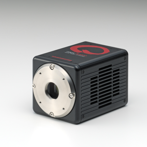
ORCA-Fusion Digital CMOS Camera
The ORCA-Fusion, built from the sensor up, balances the complex nuances of camera features to provide beautiful images and robust data at all lights levels, but especially in tough low-light conditions.
Applications & Specialities, Cameras, Products & Services, Scientific Imaging, Hamamatsu Photonics, Microscopy & Imaging
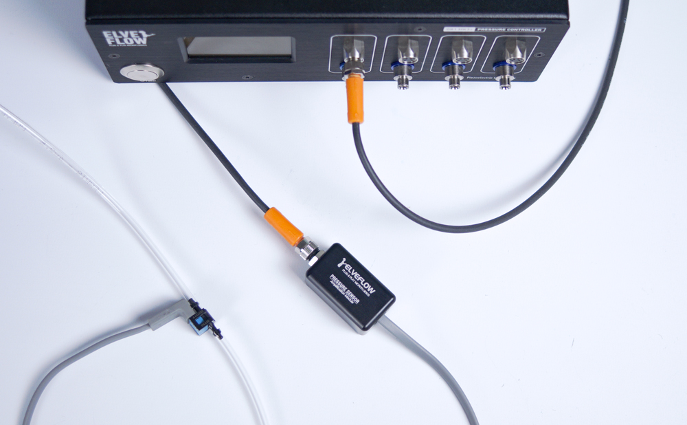
MICROFLUIDIC PRESSURE SENSOR
Elveflow provides high accuracy pressure sensors suited to liquids and gas. Its unique design has been optimised to remove dead volume and to have a low internal volume which makes it a perfect choice for microfluidic applications.
Applications & Specialities, Microfluidics, Microfluidics & Microfabrication, Products & Services, Elveflow, Microscopy & Imaging
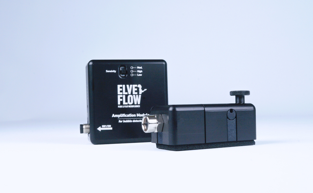
MICROFLUIDIC BUBBLE DETECTOR
The unique Inline Fluid Sensor that can detect fluid interface changes inside a clear microfluidic tubing.It can be used as a bubble detector, sensing and counting gas bubbles inside a liquid flow, or as a safety device for detecting when fluid runs out in a microfluidic line.
Applications & Specialities, Microfluidics, Microfluidics & Microfabrication, Products & Services, Elveflow, Microscopy & Imaging
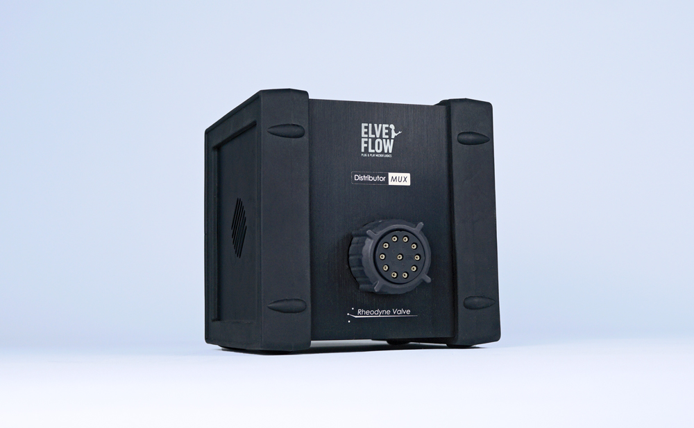
Mux Distributor – Distribution Valve
10-1 bidirectional microfluidic valve. The MUX Distributor valve is a bidirectional 10 position/11-port valve which can be used to sequentially inject or select up to 10 different fluids.
Applications & Specialities, Microfluidics, Microfluidics & Microfabrication, Products & Services, Elveflow, Microscopy & Imaging
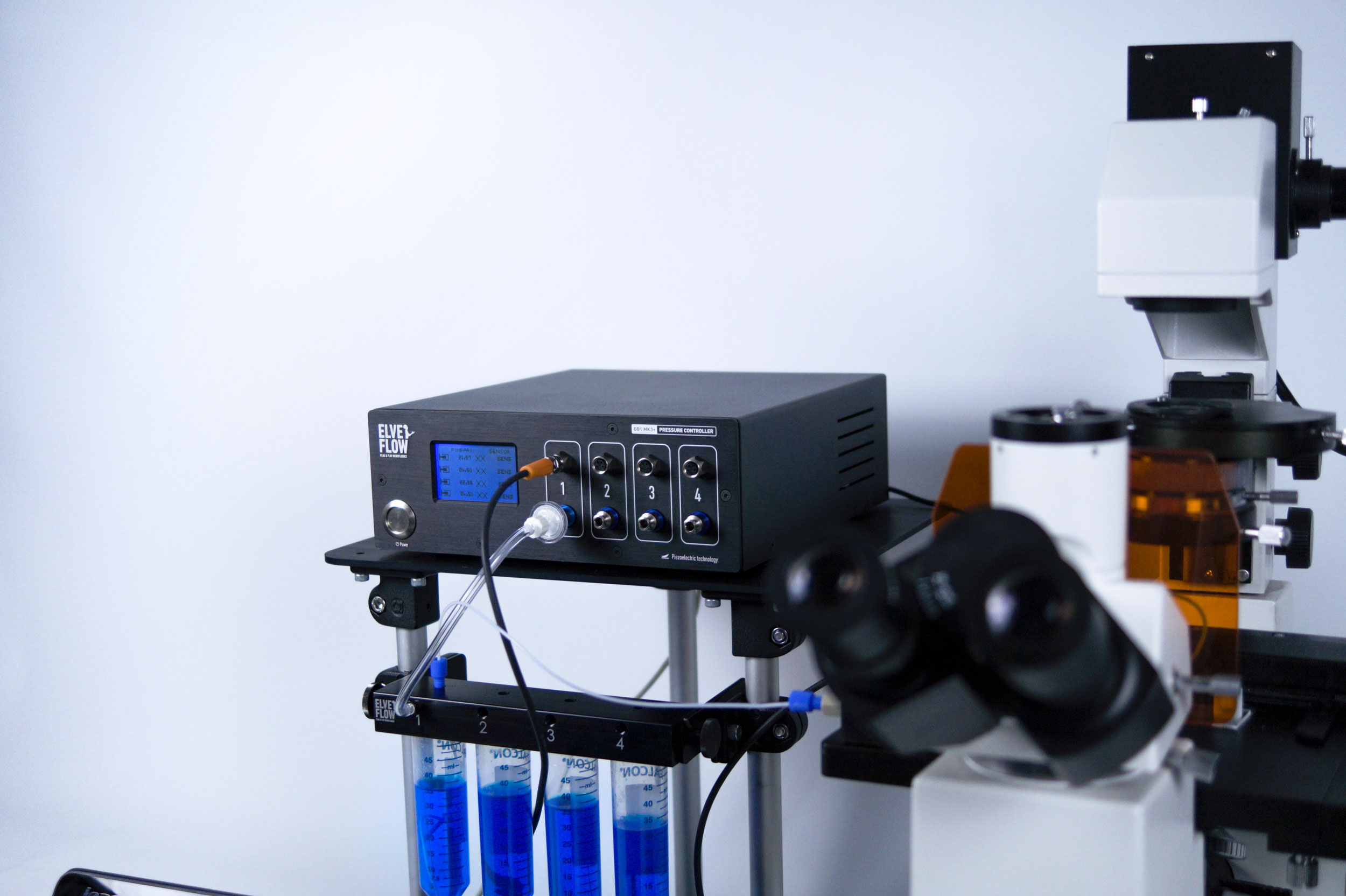
Microfluidic flow controller – OB1 MK3+
Multi-channel pressure & vacuum controller for microfluidics. The OB1 MK3+ by Elveflow is the only microfluidic flow control system in the world to use piezoelectric regulators, enabling a flow control that is 20 times more precise and 10 times faster than the other flow controllers on the market. It is 10 times more stable and up to 10 times faster than other microfluidic flow controllers.
Applications & Specialities, Microfluidics, Microfluidics & Microfabrication, Products & Services, Elveflow, Microscopy & Imaging
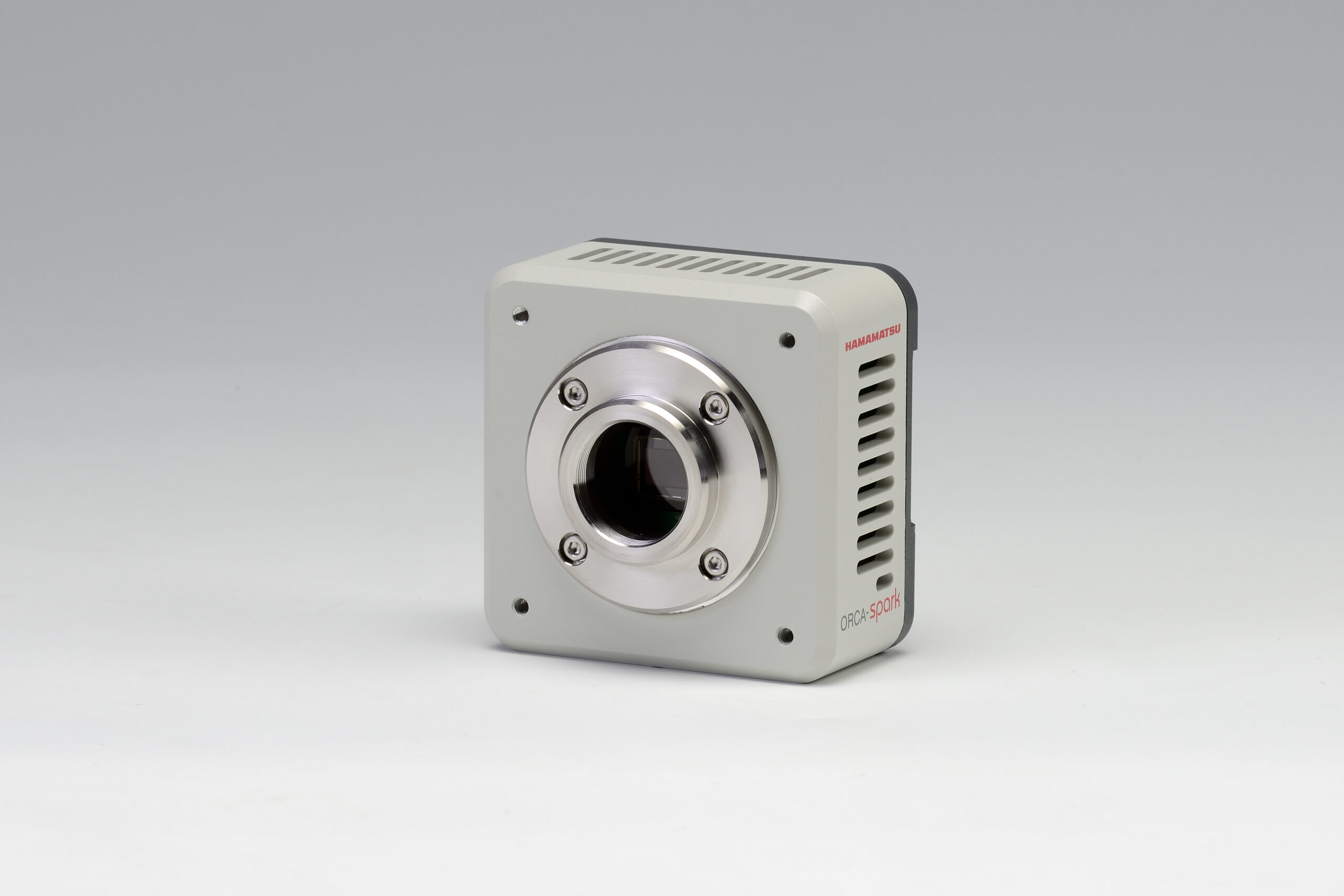
ORCA-spark Digital CMOS camera: C11440-36U
A high-sensitivity digital CMOS camera employing a 2.3 megapixel CMOS sensor comprises the ORCA-spark. Its global shutter meant to attain a high-speed readout of 65 frames/s thereby make it ideal for imaging fast-moving objects. The ORCA-spark promises to deliver readout noise levels as low was 6.6 electrons, further facilitates imaging with high S/N ratio while capturing images of dark objects.
Cameras, Instruments, Products & Services, Scientific Imaging, Microscopy & Imaging
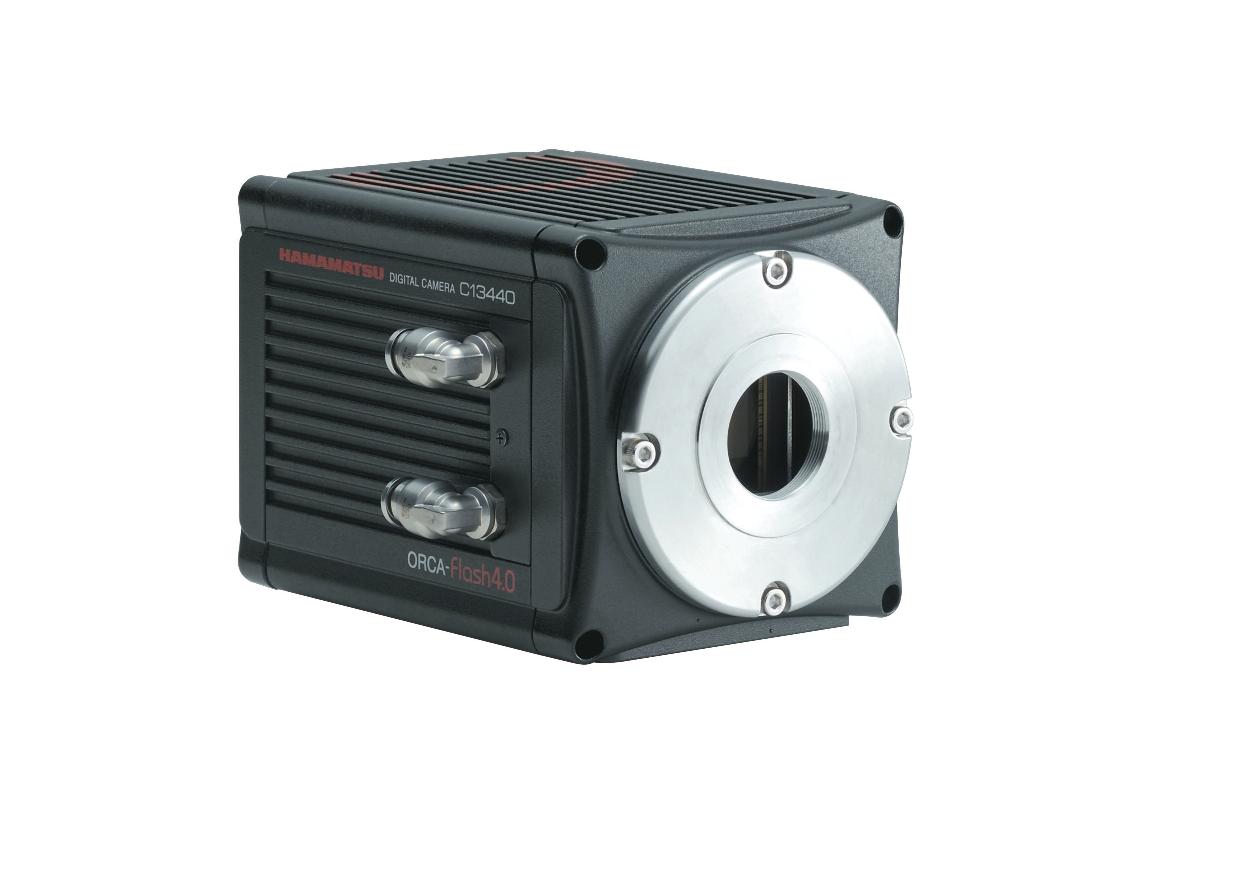
ORCA-Flash4.0 V3 Digital CMOS camera: C13440-20CU
Introducing the new ORCA-Flash4.0 V3 by Hamamatsu; developed based on their expanded experience with the advanced imaging applications along with high-performance scientific cameras. This specific camera is an expert at handling applications varying from acquiring beautiful scientific images, to experimenting detection, quantification and speed.
Cameras, Instruments, Scientific Imaging, Hamamatsu Photonics, Microscopy & Imaging
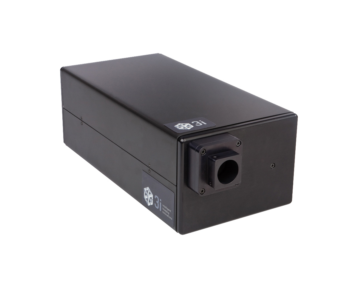
3i Ablate!
Ablate!™ laser ablation system is an attenuatable 532nm pulse laser (>60µJ pulses at 200Hz) that inflicts localized damage to a diffraction-limited spot in intracellular structures for precise laser ablation and laser wounding.
Accessories, Microscope Accessories, Photomanipulation, Products & Services, 3i, Microscopy & Imaging
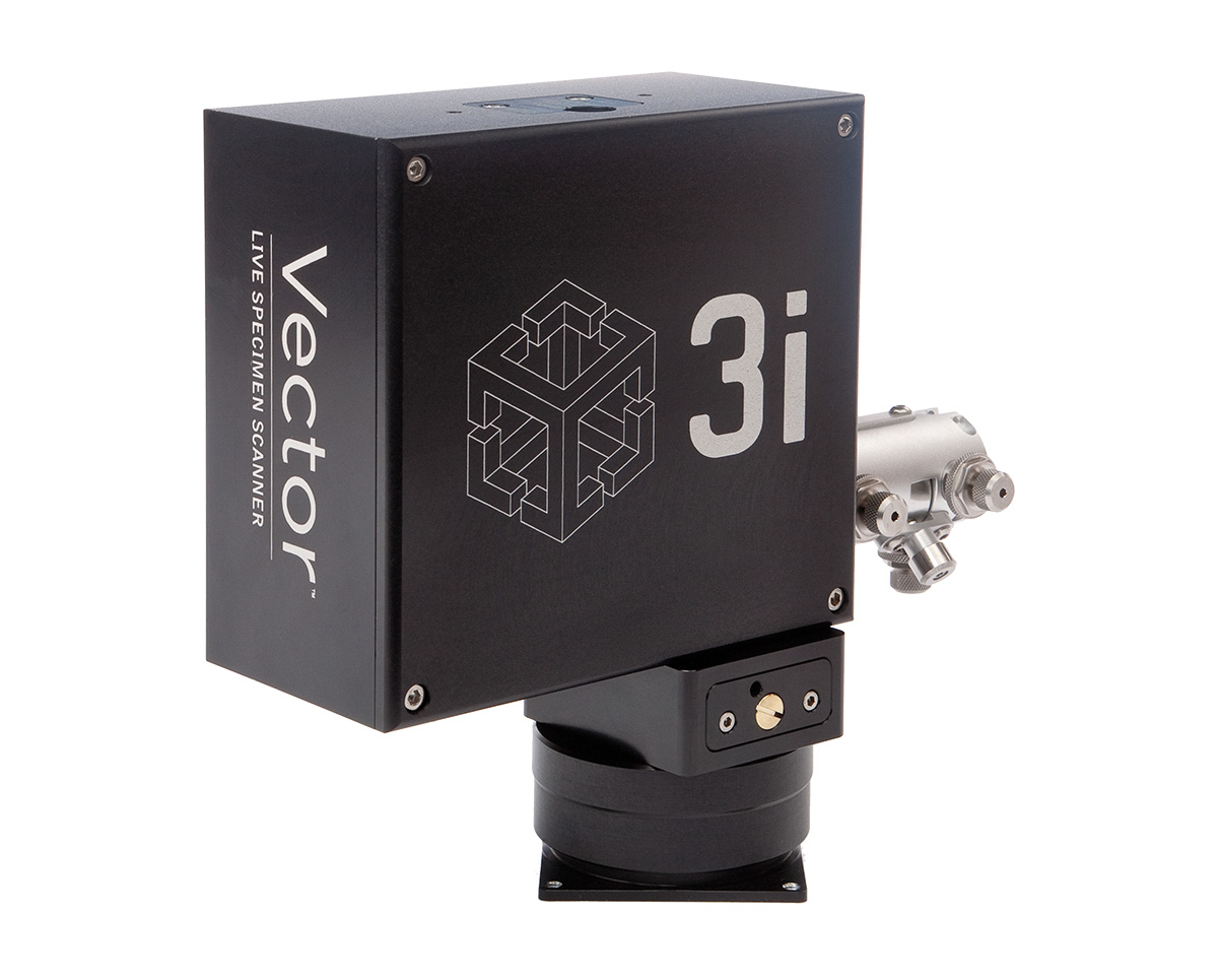
3i Vector
Vector™ is a diffraction-limited high speed X,Y scanner that accepts lasers from UV to IR.
Accessories, Applications & Specialities, Microscope Accessories, Photomanipulation, Products & Services, 3i, Microscopy & Imaging
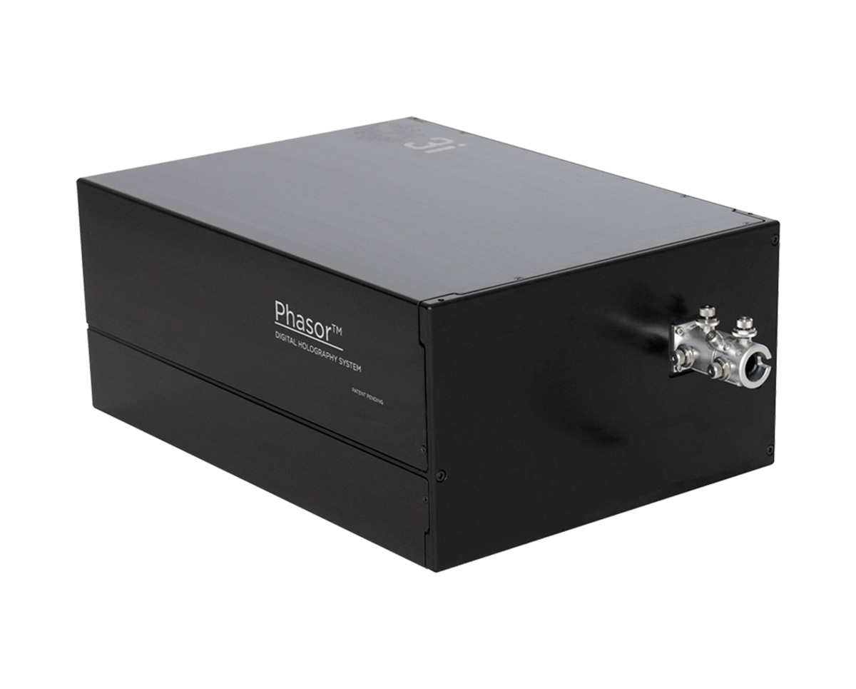
3I Phasor
Phasor is compact and modular, allowing direct attachment to a microscope imaging port. Illumination is provided by the 3i LaserStack™ through a fiber optic coupling.
Accessories, Microscope Accessories, Photomanipulation, Products & Services, 3i, Microscopy & Imaging
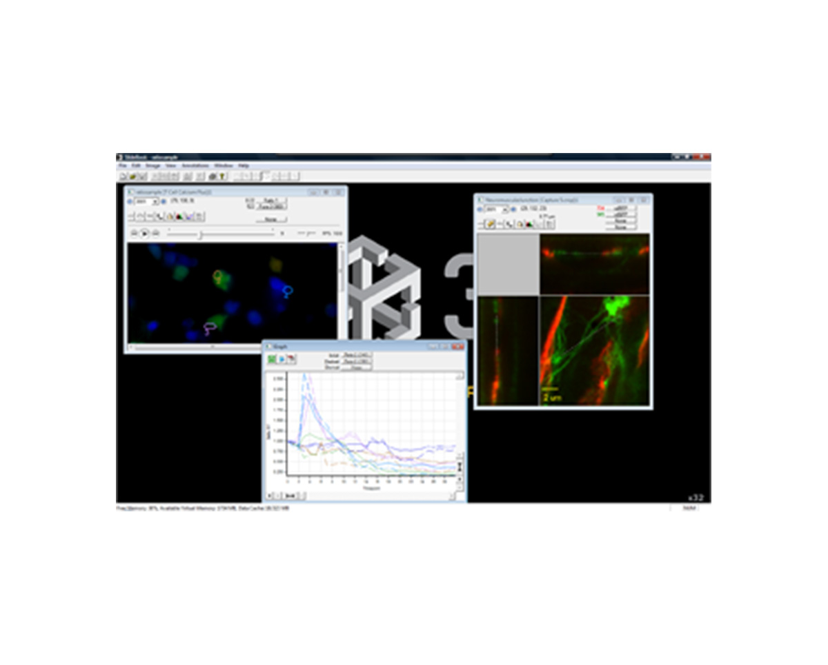
3i SlideBook
SlideBook digital microscopy software advances research microscopy through the entire experimental process.
Applications & Specialities, Imaging Software, Instruments, Microscopy, Products & Services, Scientific Imaging, Software, 3i, Microscopy & Imaging
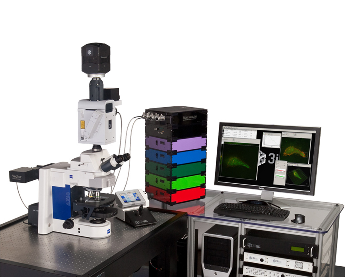
3i Everest
Everest is an upright digital microscopy workstation for all widefield fluorescence applications as well as transmitted illumination applications.
Applications & Specialities, Instruments, Microscopes, Microscopy, Products & Services, 3i, Microscopy & Imaging
Testimonials & Reviews
Dr. (Prof.), Nitesh Mohan
Professor & Head, Department of Pathology, RMCH Bareilly
DSS's expertise, dedication, and professionalism were outstanding in making the Karyotyping & FISH workshop a great success. Their knowledge and valuable insights empowered all the participants with practical skills, receiving highly positive feedback from both students as well as faculty members.

Dr. Chhaya Chande, Professor & HOD, Microbiology
GGMCJJ Hospitals, Mumbai
“Ms. Megha Dhumal (Assistant Manager- Application) has done a satisfactory demonstration of the running of the Abbott Sample preparation machine model m2000sp and the Abbott RT-PCR machine model m2000rt. We appreciate the effort made by the DSS team under these difficult conditions to help our lab to carry out the imperative Covid-19 tests.”
Dr Sunil K Arora, Professor, Deptt of Immunopathology
PGIMER, Chandigarh
“We are using Confocal Microscope and one Fluorescence Microscope. Both are working fine. The after sales services by DSS have been excellent for functioning & upkeep of the microscopes. The applications support by experts from DSS is very useful. Keep it up!”
Dr Pramod Kumar Bajaj
MD, Spermprocessor Pvt Ltd
“Really excited to see the DSS Pathology solutions exhibition booth at APCON 2019 along with Magnus. We think all the upcoming technology had been displayed along with your efforts to make it Indigenous (Made in India) is highly appreciated. Wish you all the best. Keep it up!”
Dr. Sreejesh S, Associate Professor, Dept of Hematology
PGIMER, Chandigarh
“My experience with DSS so far has been very good till now. We are getting good support in both purchase as well as in troubleshooting. Very good experience with Mr Arun, Mr Manoj, Mr Mahesh and all others from the DSS team.”
Dr Sudha S Murthy, Department of Pathology and Laboratory Medicine
BIACH & RI, Hyderabad
“I am happy with DSS and associated with 19 years and use Dako antibody. Happy with Supply but need improvement.”
Dr S Radhika MD, PhD
Professor, Deptt. Of Cytology & Gynaec Pathology, PGIMER, Chandigarh
“PGI Cytology Dept. has had a long association with DSS- Olympus Microscopy Division. They have provided excellent services- after sales service. The product is also of very good quality. We have had no problems with their products and services are of very good quality.”
Dr Nuzhat Husain
RMLIMS, Lucknow
“Have been using Dako Reagents and Dako antibodies for a while. Services and products have been good and timely.”
Dr Minu Singh
Assistant Professor, PGIMER, Chandigarh
“MRC Holland MLPA products provided by DSS are of good quality, have never faced any quality issues with their product or shipping condition. They provide prompt response upon any query.”
Mr. Krishnani Professor, SGPGI, Lucknow
“My experience with DSS so far has been excellent for the last 30 years- sales and service experience. Microscope products are very useful and sturdy with high precision.”