DSS: Redefining Biotechnology & Life Science in India


Home > Applications & Specialities > Microscopy > Scientific Imaging
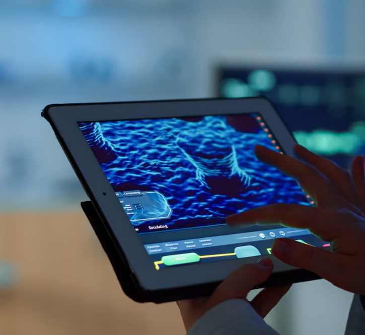
Scientific Imaging
Scientific imaging refers to the acquisition, processing, and analysis of images in a scientific context. In the field of microscopy, scientific imaging involves capturing high-quality images of biological or material samples using a microscope, and then using various imaging techniques and software tools to analyze and interpret the data.
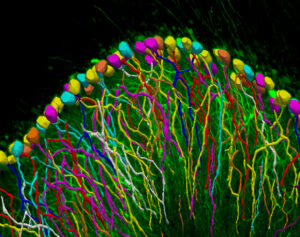
Scientific imaging in microscopy can include a range of techniques, such as brightfield imaging, fluorescence imaging, confocal microscopy, and electron microscopy. Each technique has its own strengths and limitations, and scientists may choose different techniques depending on their specific research questions and the characteristics of the sample they are studying.
In addition to capturing images, scientific imaging in microscopy often involves image processing and analysis to extract quantitative information from the data. This can involve techniques such as image segmentation, feature extraction, and image registration. The resulting data can then be used to generate visualizations, statistical analyses, and other outputs to help scientists better understand the structure and function of the sample they are studying.
In addition to capturing images, scientific imaging in microscopy often involves image processing and analysis to extract quantitative information from the data. This can involve techniques such as image segmentation, feature extraction, and image registration. The resulting data can then be used to generate visualizations, statistical analyses, and other outputs to help scientists better understand the structure and function of the sample they are studying.
SpinSR IXplore
The SpinSR offers advanced super resolution imaging for live cell studies, combining speed, precision, and efficiency in one flexible system. Designed for seamless switching between super resolution, confocal, and widefield imaging, the system captures intricate cellular structures with 120 nm resolution, enabling researchers to explore dynamic biological processes effortlessly. Advanced high-speed data processing algorithms, enabling […]
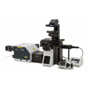
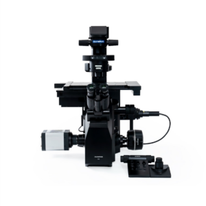
IXplore™ IX85 SPIN
The IXplore™ IX85 platform offers unparalleled customizability, enabling you to create a high-performance imaging system tailored to your unique needs. Featuring an industry-leading field number (FN) of 26.5mm, along with advanced imaging and workflow capabilities, the IXplore™ IX85 ensures superior clarity and efficiency. Capture more details with an expansive field of view (FOV) and innovative […]
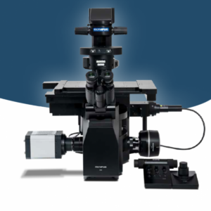
IXplore IX85 Live
Experience the next level of precision and flexibility in live cell imaging with the IXplore™ IX85 Live Inverted Microscope. Engineered to meet the rigorous demands of modern cell biology, this high-end research system delivers exceptional imaging performance with intelligent automation and robust stability. The IX85 Live system is built on a modular platform, allowing seamless […]
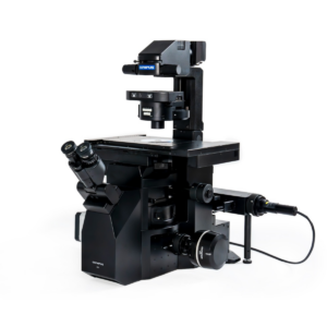
IXplore IX85 PRO
Experience advanced live-cell imaging with the IXplore IX85 Pro, a high-performance inverted microscope designed to streamline and elevate your research. Engineered for seamless automation and flexibility, this microscope system supports a wide range of imaging techniques—making it the ideal choice for complex, time-lapse, and multi-dimensional experiments. Built on optical platform, the IX85 Pro delivers exceptional […]
IX83 SpinSR
The IXplore IX83 SpinSR is a high-performance super-resolution imaging system built on the robust IX83 inverted microscope platform. Designed for researchers who demand exceptional image clarity and live-cell imaging capabilities, this system achieves resolution down to 120 nm—bridging the gap between conventional microscopy and electron microscopy.
Neurolucida
Neurolucida is a powerful analysis tools bring comprehensive quantitative morphometry to your lab, providing accuracy, efficiency, value, and results in a versatile sytem that can handle your research needs.
Stereo Investigator
Stereo Investigator is designed to help you obtain the most efficient, precise, and unbiased estimate of cell populations, as well as morphometric properties of biological structures.
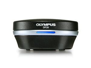
DP28
Microscope Camera for Conferencing, Teaching, and Clinical Research: The DP28 digital microscope camera combines powerful features, precise color accuracy, and 4K resolution across a wide field of view to provide stunning images for conferencing, teaching, and clinical research. With smart features, the camera eases and accelerates your microscopy tasks while delivering high image quality.
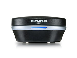
DP23
Laboratory Microscope Camera: Designed for routine life science and clinical research microscopy imaging, the DP23 digital microscope camera’s combination of smart features and good color reproduction provides high-quality images in an easy-to-use camera.
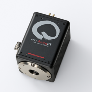
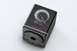
ORCA-Fusion BT Digital CMOS camera
The ORCA-Fusion BT camera is the pinnacle of scientific CMOS (sCMOS) performance. The specifications are without compromise: ultra-low readout noise, CCD-like uniformity, fast frame rates and back-thinned enabled high QE.
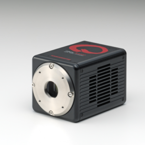
ORCA-Fusion Digital CMOS Camera
The ORCA-Fusion, built from the sensor up, balances the complex nuances of camera features to provide beautiful images and robust data at all lights levels, but especially in tough low-light conditions.
ORCA-spark Digital CMOS camera: C11440-36U
A high-sensitivity digital CMOS camera employing a 2.3 megapixel CMOS sensor comprises the ORCA-spark. Its global shutter meant to attain a high-speed readout of 65 frames/s thereby make it ideal for imaging fast-moving objects. The ORCA-spark promises to deliver readout noise levels as low was 6.6 electrons, further facilitates imaging with high S/N ratio while capturing images of dark objects.
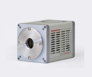
ORCA-Flash4.0 V3 Digital CMOS camera: C13440-20CU
Introducing the new ORCA-Flash4.0 V3 by Hamamatsu; developed based on their expanded experience with the advanced imaging applications along with high-performance scientific cameras. This specific camera is an expert at handling applications varying from acquiring beautiful scientific images, to experimenting detection, quantification and speed.
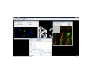
3i SlideBook
SlideBook digital microscopy software advances research microscopy through the entire experimental process.
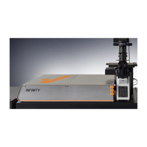
INFINITY
The INFINITY platform stands as the ultimate customisable solution for all microscopy applications. INFINITY is designed to meet every research challenge from STED and confocal microscopy to intravital imaging, material science, and optical trapping. Simply share your requirements, and we'll deliver a tailored, continuously upgradeable system precisely configured to support your research objectives.
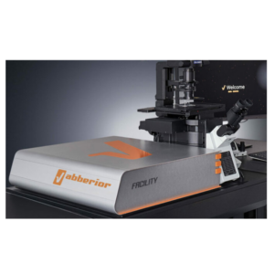
FACILITY
FACILITY offers a revolutionary combination of advanced microscopy technology and user-friendly design, setting new standards for high-performance imaging. This system integrates state-of-the-art features ideal for both confocal and super-resolution imaging, all with intuitive, effortless operation.
Abberior STAR
Our abberior STAR dyes are designed to set a new benchmark in fluorescence light microscopy, offering unparalleled performance. These dyes boast extraordinary photostability and remarkable brightness, ensuring crisp, high-resolution imaging every time. Whether you are conducting STED or confocal imaging, our STAR dyes are the ultimate choice for achieving exceptional clarity and long-lasting fluorescence, making them a vital component of your research and imaging needs. There's no better way to elevate your microscopy results.

Abberior LIVE
Experience superior live-cell imaging with abberior LIVE dyes, meticulously crafted for STED (Stimulated Emission Depletion) and confocal microscopy. These innovative dyes set new benchmarks in live-cell labeling by delivering exceptional brightness and unmatched photostability. They simplify your research workflow, ensuring high-quality results every time.
Abberior FLUX
Our abberior FLUX dyes are specifically designed to provide unparalleled performance in MINFLUX imaging. Combining outstanding photostability with exceptional brightness, these dyes enable clear and accurate visualization while minimizing background noise. Their photoswitching kinetics are finely tuned for optimal results in single-molecule localization microscopy, ensuring precise and reliable data.

Abberior CAGE
abberior CAGE dyes represent a sophisticated family of fluorophores specifically engineered for advanced imaging techniques such as PALM, STORM, and GSD microscopy. These dyes exhibit a unique property: they remain non-fluorescent until activated by UV light. Upon exposure, they are transformed into intensely bright organic probes, delivering exceptional fluorescence with remarkable precision for localization. This uncaging mechanism makes them invaluable for applications requiring the highest levels of accuracy and brightness in single-molecule and super-resolution microscopy, significantly enhancing imaging outcomes and data reliability.
Abberior Supplies
Ready-to-Image Alignment Sets - abberior ALIGNMENT SETS simplify the calibration and testing of fluorescence microscopes. Each set includes multiple microscope slides pre-prepared with nanoparticles or cell specimens, allowing for immediate imaging and precise adjustments.
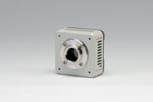
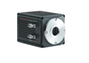
ORCA-Flash4.0 LT3 Digital CMOS camera(C11440-42U40)
The ORCA®-Flash4.0 LT3 is an advanced scientific CMOS camera specifically designed for fluorescence imaging. Designed on the success of the original ORCA®-Flash 4.0 introduced in 2011, this new model delivers exceptional performance for life science applications. It is suitable for both fundamental research and integration into diverse equipment types.
It offers an impressive readout speed of 40 frames per second, surpassing the conventional ORCA®-Flash4.0 LT+ which operates at 30 frames per second. This enhanced speed is particularly beneficial for applications requiring real-time measurements with high temporal resolution.
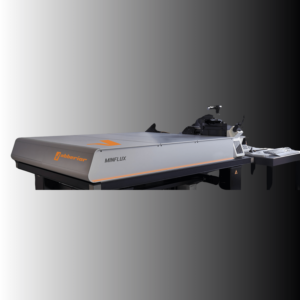
MINFLUX
The MINFLUX platform presents an unparalleled spectrum of imaging capabilities, enabling the resolution of molecular-scale structures in all three spatial dimensions. This unparalleled resolution capability, coupled with unprecedented imaging speed, unveils hitherto unobservable sample intricacies. The MINFLUX stands as the preeminent fluorescence microscope worldwide, demonstrating the highest level of power and performance in its class.
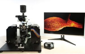
Alpha3 Light Sheet Microscope
The Alpha3, a product developed by PhaseView, has been designed to provide researchers with a versatile and high-performance light sheet microscopy solution. As an advanced fluorescence microscope, the Alpha3 incorporates intelligent optical focus sweeping capabilities to ensure uniform and artifact-free imaging across the entirety of the field of view.
SLIDEVIEW DX VS-M1
By integrating our state-of-the-art technologies, such as X Line objectives and True Color LED illumination, we have developed a rapid slide scanner capable of producing high-resolution images that match the quality of those seen through a microscope. The SLIDEVIEW™ DX whole slide imaging system incorporates advanced technology that enhances the digital pathology workflow, enabling pathologists to efficiently diagnose cases with dependable, top-notch images.
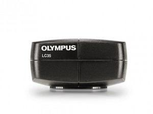
LC35
The LC35 digital microscope camera offsets picture quality with reasonableness to convey a great value for standard brightfield imaging applications.
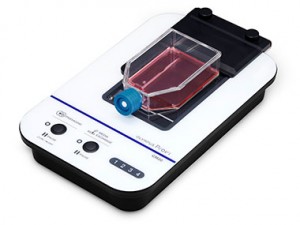
CM30
Formulating cell cultures can be an expensive, complicated, and time-consuming procedure.
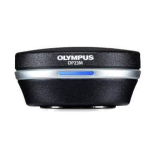
DP23M
DP23M camera makes it simple to quickly obtain high-quality fluorescence images required for sample inspection and routine fluorescence imaging. It is a monochrome digital microscope camera.
ORCA-Quest qCMOS camera (C15550-20UP)
The camera achieves the ultimate in quantitative imaging.
Testimonials & Reviews

Dr. (Prof.), Nitesh Mohan
Professor & Head, Department of Pathology, RMCH Bareilly
DSS's expertise, dedication, and professionalism were outstanding in making the Karyotyping & FISH workshop a great success. Their knowledge and valuable insights empowered all the participants with practical skills, receiving highly positive feedback from both students as well as faculty members.

Dr. Chhaya Chande, Professor & HOD, Microbiology
GGMCJJ Hospitals, Mumbai
“Ms. Megha Dhumal (Assistant Manager- Application) has done a satisfactory demonstration of the running of the Abbott Sample preparation machine model m2000sp and the Abbott RT-PCR machine model m2000rt. We appreciate the effort made by the DSS team under these difficult conditions to help our lab to carry out the imperative Covid-19 tests.”
Dr Sunil K Arora, Professor, Deptt of Immunopathology
PGIMER, Chandigarh
“We are using Confocal Microscope and one Fluorescence Microscope. Both are working fine. The after sales services by DSS have been excellent for functioning & upkeep of the microscopes. The applications support by experts from DSS is very useful. Keep it up!”
Dr Pramod Kumar Bajaj
MD, Spermprocessor Pvt Ltd
“Really excited to see the DSS Pathology solutions exhibition booth at APCON 2019 along with Magnus. We think all the upcoming technology had been displayed along with your efforts to make it Indigenous (Made in India) is highly appreciated. Wish you all the best. Keep it up!”
Dr. Sreejesh S, Associate Professor, Dept of Hematology
PGIMER, Chandigarh
“My experience with DSS so far has been very good till now. We are getting good support in both purchase as well as in troubleshooting. Very good experience with Mr Arun, Mr Manoj, Mr Mahesh and all others from the DSS team.”
Dr Sudha S Murthy, Department of Pathology and Laboratory Medicine
BIACH & RI, Hyderabad
“I am happy with DSS and associated with 19 years and use Dako antibody. Happy with Supply but need improvement.”
Dr S Radhika MD, PhD
Professor, Deptt. Of Cytology & Gynaec Pathology, PGIMER, Chandigarh
“PGI Cytology Dept. has had a long association with DSS- Olympus Microscopy Division. They have provided excellent services- after sales service. The product is also of very good quality. We have had no problems with their products and services are of very good quality.”
Dr Nuzhat Husain
RMLIMS, Lucknow
“Have been using Dako Reagents and Dako antibodies for a while. Services and products have been good and timely.”
Dr Minu Singh
Assistant Professor, PGIMER, Chandigarh
“MRC Holland MLPA products provided by DSS are of good quality, have never faced any quality issues with their product or shipping condition. They provide prompt response upon any query.”
Mr. Krishnani Professor, SGPGI, Lucknow
“My experience with DSS so far has been excellent for the last 30 years- sales and service experience. Microscope products are very useful and sturdy with high precision.”