DSS: Redefining Biotechnology & Life Science in India




























Home > Products & Services > Instruments > Microscopes
Microscopes
A microscope is an instrument that magnifies objects, too small to be seen with an unaided eye, producing larger images using a combination of lenses. The student lab microscope in general is a brightfield microscope, implying that visible light is passed through the sample and used to shape a picture directly, without any changes. Another sort of light microscopy is fluorescence microscopy, which is utilized for image samples that fluoresce (absorb one frequency of light and emanate another). For additional improved resolution, Confocal Microscopy is utilized. A confocal microscope utilizes a laser to energize a thin layer of the sample and collects just the transmitted light coming from the target layer, delivering a sharp image without interference from fluorescent particles in the surrounding layer. The selection of the type of microscopes is based on the type of sample (size, thickness, dynamics, physiology etc.) that needs to be viewed or analyzed.


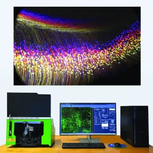
SLICE – Light Sheet Microscope
SLICE – The New Standard for Accessible Light Sheet Imaging SLICE™: a paradigm shift in light-sheet microscopy that combines high performance with unprecedented affordability and a compact device footprint. Invented by leading researchers at Columbia University, SLICE delivers advanced 3D imaging capabilities to research labs of all sizes. This innovative system offers high-resolution imaging of […]
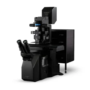
Evident FLUOVIEW™ FV5000 Confocal Microscope
The FLUOVIEW™ FV5000 from Evident elevates scientific imaging with unmatched clarity, speed, and quantitative accuracy. Built on a century of optical excellence and advanced further by Evident innovation, the FV5000 delivers superior performance from surface-level structures to deep-tissue imaging in thick, living samples. Engineered for researchers who demand precision, flexibility, and reproducibility, this next-generation platform […]

Evident BX51WI Fixed-Stage Upright Microscope
The Olympus BX51WI is a high-performance upright microscope meticulously designed for advanced in vivo and in vitro electrophysiological research. Engineered to meet the exacting demands of patch-clamp experiments, it offers exceptional stability and accessibility, with a slim body and elevated frame that provide ample space for micromanipulators. Its modular design and compatibility with long working […]

Evident CX33 Biological Microscope
Designed to streamline everyday laboratory routines, the Olympus CX33 delivers clear, bright images with minimal effort. It features a comfortable ergonomic build that allows users to operate the microscope without changing their natural hand positions, reducing strain during long hours of observation. Its LED light source provides reliable, color-accurate illumination with minimal maintenance, and the […]

Evident BX53 Upright Microscope
The BX53 is a versatile system microscope that can be configured to meet virtually any research need. It supports a wide range of fluorescence imaging applications, and has a range of advanced features for enhanced operating ease and process flexibility.
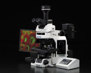
Evident BX63 Microscope
The BX63 offers outstanding stability and imaging precision for research applications, with the convenience of a touch screen interface for easy operation. Additionally, it features a detachable controller that can be positioned to suit the operator's preference or workflow requirements.

Evident MVX10 Microscope
The MVX10 MacroView is a research Macro Zoom Fluorescence Microscope. The MVX10 addresses the tradeoffs between stereo and compound imaging systems, offering high numerical apertures from a compound microscope along with long working distances and larger fields of view from a stereo microscope.

Evident SZX16 Microscope
The new SZX16 is designed for advanced research and has a maximum numerical aperture of 0.30, producing a resolution of 900 line pairs per millimeter.

Evident SZX10 Microscope
The Evident SZX10 is designed for routine research. It offers a maximum numerical aperture (NA) of 0.2, producing a resolution of 600 line pairs per millimetre.

Evident IX73 Inverted Microscope
The IX73 inverted microscope system sets new standards in advanced live cell imaging with its stable compact frame, outstanding optical performance and maximum flexibility.

Evident IX83 Microscope
The fully motorised and automated IX83 is the most advanced member of the IX3 series of imaging systems for life science researchers and offers a new level of usability and application flexibility.
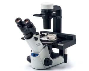
Evident CKX53 Microscope
The Evident CKX53 microscope delivers excellent performance, with improved image quality and ergonomics, as well as a comfortable workflow for a variety of cell culture needs such as live cell observation, cell sampling and handling, fluorescence observation, and image capture.
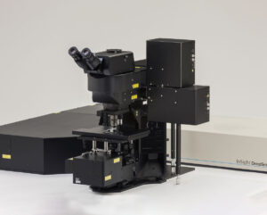
Evident FVMPE-RS Multiphoton Microscope
With the Olympus FVMPE-RS, countless possibilities for deep tissue observation are finally realized. The system delivers unmatched high-speed imaging, essential for capturing the dynamic in vivo response, with fine laser control pinpointing the precise site for optimum excitation efficiency - even deep within the sample.
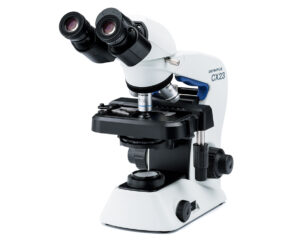
Evident CX23 Microscope
The CX23 microscope unique features accommodates the student and every requirement in the educational setting. This cost-effective system provides easy and safe operation throughout the workflow and ensures outstanding optical performance with large field number (FN) 20
Evident BX46 Clinical Microscope
The Olympus new BX46 defines a revolutionary new standard in laboratory and clinical micrscopy.
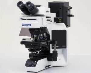
Evident BX43 Clinical Microscope
The BX43 supports brightfield, phase contrast, simple polarized light, and darkfield observation. Its Ergonomic design can provide enhanced operator comfort during long hours of use.
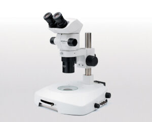
Evident SZX7 Microscope
In operation and optical performance,the SZx7 is designed for ease of use and working comfort in every task,from routine preparations to advanced research.
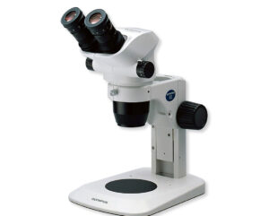
Evident SZ61TR Stereo Zoom Microscope
The SZ61TR zoom stereo microscope is ideal for routine and advanced stereo microscopic observation in life science, especially for those applications that require image documentation.
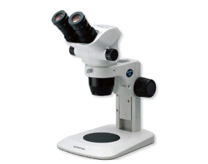
Evident SZ51 Stereo Microscope
The versatile and cost-efficient SZ51 zoom stereo microscope offers a broad range of functions for observation and documentation of specimen in life science.
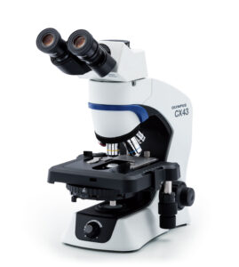
Evident CX43 Microscope
The CX43 microscope enable users to remain comfortable during long periods of routine microscopy observations.
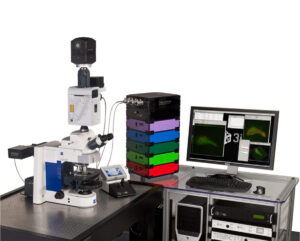
3i Everest
Everest is an upright digital microscopy workstation for all widefield fluorescence applications as well as transmitted illumination applications.
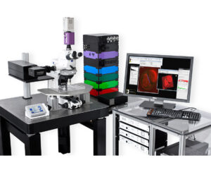
3i VIVO
VIVO incorporates advanced optics, cameras, computers and proprietary electronics to achieve unparalleled speed, precision and flexibility in live tissue image acquisition.
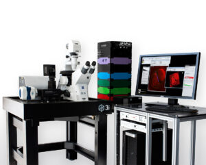
3i Marianas
Marianas incorporates advanced optics, cameras, computers and proprietary electronics to achieve unparalleled speed, precision and flexibility in live cell image acquisition.
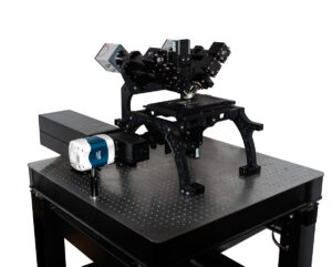
3i diSPIM
Dual-view Inverted Selective Plane Illumination (diSPIM) is a light sheet method offering low phototoxicity in 3D imaging making it possible to image living small organisms over extended periods of time.
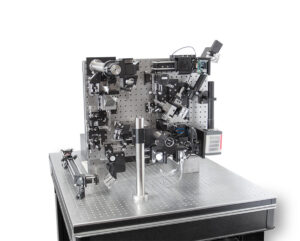
3i Lattice LightSheet
Lattice LightSheet uses ultra-thin sheets of light to image 3D cellular dynamics for hundreds of volumes at dozens of frames per second at diffraction-limited resolution and super-resolution.
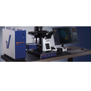
STEDYCON 2
The STEDYCON transforms your existing widefield microscope into an advanced confocal and STED nanoscope, achieving a remarkable resolution down to 30 nanometers. With just a free camera port and a quality objective lens, you can enhance your imaging capabilities significantly.
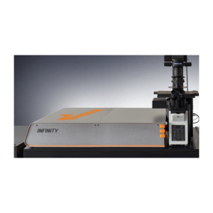
INFINITY
The INFINITY platform stands as the ultimate customisable solution for all microscopy applications. INFINITY is designed to meet every research challenge from STED and confocal microscopy to intravital imaging, material science, and optical trapping. Simply share your requirements, and we'll deliver a tailored, continuously upgradeable system precisely configured to support your research objectives.
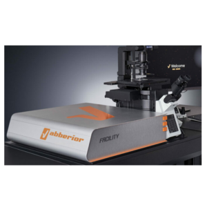
FACILITY
FACILITY offers a revolutionary combination of advanced microscopy technology and user-friendly design, setting new standards for high-performance imaging. This system integrates state-of-the-art features ideal for both confocal and super-resolution imaging, all with intuitive, effortless operation.
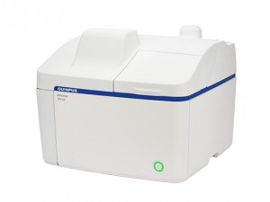
Evident APEXVIEW™ APX100 Digital Imaging System
Made with well-known Olympus optics, an reflexive user interface, a powerful AI, and a suite of smart features, the APX100 system combines ease of use with high-quality image data to fit your research needs.
Laboratory Microscopes for Research, Clinical and Educational Applications
A microscope lets you see, document and analyse things too small for the naked eye. Beyond the optics, most current systems add illumination control, cameras, motorised components and software — the goal is repeatable observation, not just a bigger image.
A pathology lab, a neuroscience lab and a teaching lab are not shopping for the same microscope. Sample type and thickness matter, sure, but so do contrast technique, resolution, working distance, whether the cells are alive or fixed, and what happens to the images afterward — get any of those wrong and the "right" microscope on paper turns out to be the wrong one on the bench.
What Types of Microscopes Are Used in Laboratories?
Compound and Biological Microscopes
A compound microscope magnifies thin specimens — slides, smears, tissue sections — using an objective and eyepiece lens working together. When it's set up for biological samples specifically, people call it a biological microscope, though the two terms overlap heavily in practice. These optical microscopes are the workhorses of teaching labs, routine laboratory work and clinical observation, usually built around brightfield illumination, with phase contrast, darkfield or simple polarised light available on some models.
Stereo and Macro Observation Microscopes
Stereo microscopes give each eye its own optical path, which produces a genuine three-dimensional view at fairly low magnification. The wider field of view and longer working distance make them the right tool for larger or opaque specimens — dissection work, sample prep, inspection, documentation. Macro zoom systems sit in between: stereo-like working distance with sharper optical performance than a typical stereo scope.
Inverted Microscopes for Cell Culture
Flip the objectives underneath the stage and you get an inverted microscope — built to look up through the base of a culture dish or flask without disturbing what's inside. That's why inverted platforms dominate cell-culture observation, sampling, live-cell imaging and time-lapse work. Add fluorescence, environmental control, a motorised stage or digital capture, and the same platform stretches into more demanding research.
Fluorescence and Confocal Microscopes
Fluorescence microscopy hits a sample with specific wavelengths of light to excite fluorescent labels, making it possible to pick out cells, proteins or structures that transmitted light alone would blur together.
Confocal microscopy goes a step further: a pinhole strips out the out-of-focus light, sharpening contrast and enabling optical sectioning even in thicker specimens. This is a deep topic on its own — for the details, see DSS Imagetech's confocal microscopy systems.
Digital and Automated Imaging Systems
Pair microscope optics with a camera and acquisition software and you have a digital microscopy workflow. Add autofocus, motorised stage movement or automated capture, and it starts supporting things a manual setup can't: consistent documentation, quantitative analysis, remote review, higher throughput. Worth checking compatible microscope cameras while you're planning the configuration.
Light-Sheet and Super-Resolution Microscopy
Light-sheet microscopy illuminates only a thin plane of the sample, so the rest of it isn't sitting under unnecessary light — useful for long three-dimensional imaging of living or light-sensitive specimens.
Super-resolution pushes past the conventional diffraction limit using optical and computational tricks, aimed squarely at subcellular and molecular structure. See DSS Imagetech's super-resolution microscopy solutions for the relevant systems.
Specialised Microscopes for Electrophysiology
Patch-clamp and related electrophysiology work needs a fixed-stage upright microscope — mechanically stable, with enough clearance for micromanipulators around live cells or tissue. Browse electrophysiology systems for application-specific options.
How to Choose the Right Microscope
Start with the sample and the question you're trying to answer, not the magnification number. A higher number doesn't automatically mean more detail — resolution, numerical aperture, contrast method and sample prep all decide that.
Before shortlisting a research microscope, work through:
- Application — routine observation, teaching, clinical laboratory work, cell culture, fluorescence imaging, electrophysiology or advanced research each pull toward different systems.
- Sample type — thin slides, live cells, tissue sections, whole organisms, thick or opaque material.
- Resolution and magnification — pick objectives for the detail you actually need; resolution matters more than the magnification figure on the box.
- Contrast method — brightfield, phase contrast, darkfield, fluorescence, confocal, or something else.
- Working distance and field of view — enough room for culture vessels or manipulation tools, without losing the field you need to observe.
- Live or fixed samples — live-cell work often needs an inverted platform, environmental control and illumination that doesn't overexpose the sample.
- Image capture and analysis — camera compatibility, image quality, measurement tools, data handling, reporting.
- Manual or automated operation — motorised focus, stages and acquisition improve repeatability once workflows get complex or high-throughput.
- Practical lab requirements — bench space, training, installation and support needs, sorted before you finalise a configuration.
Comparing Common Microscope Types
| Microscope type | Typical sample | Primary advantage | Common application |
|---|---|---|---|
| Compound or biological | Slides, smears, thin tissue sections | Detailed transmitted-light observation | Teaching, routine and clinical laboratory observation |
| Stereo or macro | Whole specimens, larger opaque samples | 3D view, longer working distance | Dissection, handling, inspection, documentation |
| Inverted | Live cells in dishes or flasks | Observation through the vessel base | Cell culture, live-cell imaging |
| Fluorescence | Fluorescently labelled cells or tissue | Visualises selected labelled structures | Cell biology, fluorescence-based research |
| Confocal | Fluorescent or thicker specimens | Optical sectioning, less out-of-focus light | Cellular and three-dimensional imaging |
| Light-sheet | Living, light-sensitive specimens | Less illumination outside the focal plane | Extended three-dimensional imaging |
| Super-resolution | Subcellular structures | Detail beyond conventional light-microscopy limits | Advanced cellular and molecular research |
| Electrophysiology upright | Live cells or tissue preparations | Stable stage, room for manipulation | Patch-clamp, neuroscience research |
Where Are Laboratory Microscopes Used?
In life-science and biotechnology research, microscopy underpins the study of cells, tissues and biological processes. Cell-culture labs lean on inverted platforms to watch growth and behaviour; fluorescence, confocal, light-sheet and super-resolution systems come in when researchers need molecular specificity, optical sectioning, extended observation, or finer structural detail than a standard setup gives.
In pathology and other clinical laboratory workflows, biological and compound microscopes support trained professionals examining prepared specimens as part of established procedures — the microscope itself doesn't make the diagnosis. Universities and teaching labs care more about durability, ease of use and operator comfort than raw capability. Neuroscience and electrophysiology labs need stable upright platforms with room for electrodes and manipulation equipment.
From Routine Observation to Advanced Scientific Imaging
DSS Imagetech's range runs from routine optical observation to specialised scientific imaging. Evident CX series systems handle educational and everyday laboratory work; BX series microscopes cover clinical laboratory and research needs; Evident IX inverted platforms are built for cell-culture and live-cell applications. Confocal, multiphoton, light-sheet, digital-imaging and super-resolution platforms round out the more advanced end.
The portfolio draws on brands including Evident, 3i, ABBERIOR INSTRUMENTS and MBF Bioscience. Specifications, laboratory accessories and configurations differ by product, so it's worth checking each system against your sample, application and workflow before deciding.
Why Work With DSS Imagetech for Microscopy Requirements?
DSS Imagetech is one of the leaders in Microscopy and scientific imaging in India, from routine compound and biological microscopes through to confocal, light-sheet and super-resolution platforms. That range means you can weigh options against your actual research, clinical or educational requirement rather than settling for whatever's on the shelf.
The website also connects microscope categories to related applications, cameras and imaging systems — useful if you're planning a workflow rather than buying an isolated instrument.
Discuss Your Microscopy Requirements
When you enquire, it helps to mention the sample type, the intended application, the imaging method you expect to need, documentation or analysis requirements, how much automation you want, and any existing equipment the new system has to work with. That's enough for the DSS Imagetech team to point you toward systems worth evaluating. Use the enquiry option on the product page, or contact DSS Imagetech directly.
Frequently Asked Questions
Find answers to common questions about microscopes, their applications, and selection.
What is a laboratory microscope used for?
A laboratory microscope is used to observe and document cells, tissues, microorganisms and other specimens that cannot be examined clearly with the unaided eye. Its practical capabilities depend on the optical configuration, illumination method, camera and imaging software.
What is the difference between a compound microscope and a biological microscope?
A compound microscope magnifies a specimen through an objective-and-eyepiece system. Call that same setup a biological microscope when it's configured for cells, smears and tissue sections. Most people use the two terms interchangeably anyway.
Which microscope is suitable for viewing living cells?
Inverted microscopes, almost always — the objectives sit below the stage and view the sample from underneath, so you're not disturbing the dish. Fluorescence, confocal or light-sheet techniques might come into it too, depending on the question you're asking and how long you need to keep imaging.
Is magnification the same as resolution?
No. Magnification is how much bigger the image looks; resolution is whether you can actually tell two close details apart. Push magnification without enough optical resolution and you just get a bigger, blurrier image — not more information.
How do I choose a research microscope?
Weigh sample type, required resolution, contrast method, working distance, field of view, live-cell needs, camera and software requirements, automation level, available space and operator training together. The right configuration matches the whole imaging workflow, not just one spec.
Can laboratory microscopes capture and analyse digital images?
Yes. Many current laboratory microscopes can be paired with digital cameras and imaging software for capture, storage, measurement, analysis and sharing. What's actually possible depends on the specific microscope, camera and software combination.
Testimonials & Reviews


Dr. (Prof.), Nitesh Mohan
Professor & Head, Department of Pathology, RMCH Bareilly
DSS's expertise, dedication, and professionalism were outstanding in making the Karyotyping & FISH workshop a great success. Their knowledge and valuable insights empowered all the participants with practical skills, receiving highly positive feedback from both students as well as faculty members.

Dr. Chhaya Chande, Professor & HOD, Microbiology
GGMCJJ Hospitals, Mumbai
“Ms. Megha Dhumal (Assistant Manager- Application) has done a satisfactory demonstration of the running of the Abbott Sample preparation machine model m2000sp and the Abbott RT-PCR machine model m2000rt. We appreciate the effort made by the DSS team under these difficult conditions to help our lab to carry out the imperative Covid-19 tests.”

Dr Sunil K Arora, Professor, Deptt of Immunopathology
PGIMER, Chandigarh
“We are using Confocal Microscope and one Fluorescence Microscope. Both are working fine. The after sales services by DSS have been excellent for functioning & upkeep of the microscopes. The applications support by experts from DSS is very useful. Keep it up!”

Dr Pramod Kumar Bajaj
MD, Spermprocessor Pvt Ltd
“Really excited to see the DSS Pathology solutions exhibition booth at APCON 2019 along with Magnus. We think all the upcoming technology had been displayed along with your efforts to make it Indigenous (Made in India) is highly appreciated. Wish you all the best. Keep it up!”
Dr. Sreejesh S, Associate Professor, Dept of Hematology
PGIMER, Chandigarh
“My experience with DSS so far has been very good till now. We are getting good support in both purchase as well as in troubleshooting. Very good experience with Mr Arun, Mr Manoj, Mr Mahesh and all others from the DSS team.”

Dr Sudha S Murthy, Department of Pathology and Laboratory Medicine
BIACH & RI, Hyderabad
“I am happy with DSS and associated with 19 years and use Dako antibody. Happy with Supply but need improvement.”
Dr S Radhika MD, PhD
Professor, Deptt. Of Cytology & Gynaec Pathology, PGIMER, Chandigarh
“PGI Cytology Dept. has had a long association with DSS- Olympus Microscopy Division. They have provided excellent services- after sales service. The product is also of very good quality. We have had no problems with their products and services are of very good quality.”

Dr Nuzhat Husain
RMLIMS, Lucknow
“Have been using Dako Reagents and Dako antibodies for a while. Services and products have been good and timely.”
Dr Minu Singh
Assistant Professor, PGIMER, Chandigarh
“MRC Holland MLPA products provided by DSS are of good quality, have never faced any quality issues with their product or shipping condition. They provide prompt response upon any query.”

Mr. Krishnani Professor, SGPGI, Lucknow
“My experience with DSS so far has been excellent for the last 30 years- sales and service experience. Microscope products are very useful and sturdy with high precision.”